
Table of Contents
Introduction: The Critical Role of Advanced Diagnostics in 2025 MVA Litigation:
How Dr. Alex Jimenez Positions Plaintiff’s Attorneys for Jury Success and Maximum Monetary Recoveries
In 2025, motor vehicle accidents (MVAs) continue to be a leading cause of spinal injuries in Texas. According to statistics from the Texas Department of Transportation, there are over 500,000 crashes each year, resulting in tens of thousands of cases of spinal trauma. These incidents often lead to complex personal injury claims, in which insurance companies aggressively dispute causation by labeling acute pathologies as “pre-existing degeneration” to reduce payouts. For lawyers representing injured clients, the difficulty lies in not just proving who is at fault but also in scientifically showing that the accident caused the injury, when it happened, and how serious the disability is—these details can influence how sympathetic a jury feels and the amount of money awarded for pain, suffering, lost wages, and future medical costs.
The landscape of personal injury litigation in Texas has changed significantly, with insurance companies using advanced AI tools to dispute claims by attributing spinal issues to “pre-existing degeneration” rather than traumatic events. This change has made it harder for attorneys to obtain fair compensation for clients involved in motor vehicle crashes, workplace accidents, or slip-and-fall cases. Critical to winning claims is the ability to scientifically prove causality—demonstrating that the injury was directly caused by the incident—and to precisely date the injury to exclude chronic conditions.
 This aspect is where Dr. Alex Jimenez and the practitioners at Injury Medical Clinic PA in El Paso, Texas, stand out as the top choice for plaintiff’s counsel. As a dual-licensed Doctor of Chiropractic (DC) and board-certified Family Nurse Practitioner (APRN, FNP-BC) with over 30 years of experience, Dr. Jimenez has built his clinic into the preferred destination for accident-related injuries. His recent seminar, recorded in October 2025, highlighted why lawyers choose to work with our office: we are excellent at understanding and explaining complex MRI results, providing strong evidence of how and when injuries occurred in legal cases, and serving as reliable expert witnesses who can clearly explain injury details to juries.
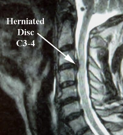
This guide uses expert information from sites that focus on diagnosing and treating injuries from motor vehicle accidents (MVAs), like the Mayo Clinic’s details on whiplash and spinal cord injuries, the Cleveland Clinic’s summaries on spinal cord injuries, and specialized orthopedic sites like Apex Spine and Neurosurgery, to back up our methods with research-based insights. For example, Mayo Clinic emphasizes that whiplash symptoms from MVAs, such as neck pain and stiffness, often begin within days, which aligns with our temporal-onset evaluations for causality. Cleveland Clinic notes that MVAs are the leading cause of spinal cord injuries, underscoring the importance of advanced imaging like MRI to detect herniated discs and edema—tools we excel at using to demonstrate impairment.
This guide delves deeply into our processes, ensuring attorneys understand how we meticulously evaluate cases to substantiate claims. We’ll illustrate various case types, justifying dual-licensed treatment in the El Paso community, and provide insightful explanations on how demystifying MRI findings for juries reveals the “invisible” disabilities from MVAs—such as chronic pain preventing work or family activities—leading to stronger cases and higher recoveries. Our goal is to instill confidence in legal counsel, positioning Injury Medical Clinic as the premier clinic for accident injuries, where science meets advocacy for plaintiff success.
Section 1: Our Practice’s Capability to Assess and Interpret Complex MRI Findings—The Foundation for Accurate Diagnosis in MVA Cases
At Injury Medical Clinic PA, advanced MRI interpretation is not just a service—it’s the cornerstone of our medico-legal expertise, enabling us to uncover subtle spinal pathologies that general practitioners might overlook. As detailed in expert sites like Mayo Clinic’s spinal cord injury diagnosis page, MRI is essential for showing trauma from MVAs, including herniated discs, vascular irregularities, bleeding, inflammation, and ligament injuries. We build on this expertise by employing a systematic multi-sequence approach to assess MVA-related spinal injuries, where high-speed impacts can cause hyperflexion, hyperextension, or compression forces leading to devastating soft tissue damage.
1.1 Understanding Spinal Anatomy and Pathophysiology in MVA Contexts—Building Jury Empathy Through Clear Explanations
To fully appreciate our MRI assessment capabilities, it’s essential to start with spinal anatomy, as we do in expert testimony to educate juries on the true extent of impairment. The spine consists of 33 vertebrae, intervertebral discs, ligaments, facet joints, and the spinal cord, all of which are vulnerable in MVAs. Discs, with their annulus fibrosus (outer fibrous ring) and nucleus pulposus (inner gel-like core), act as shock absorbers. In a rear-end collision, for example, the sudden force can tear the annulus, extrude the nucleus, and compress nerve roots, resulting in radiculopathy—shooting pain down the arms or legs that impairs daily activities like driving or lifting objects.
Expert sites like the National Institute of Neurological Disorders and Stroke (NINDS) highlight that MVAs are a primary cause of spinal cord injuries (SCI), often leading to paralysis or sensory loss below the injury site. The epidural venous plexus, or Batson’s plexus—a valveless network of veins in the epidural space—can engorge due to pressure changes in trauma, mimicking disc herniations on MRI and causing neural compression. As per NINDS, these conditions can facilitate complications like bleeding or inflammation, which we differentiate using contrast-enhanced imaging.
Dural tenting, a phenomenon in which disc protrusions create negative-pressure spaces filled by compliant veins, adds diagnostic complexity. The vacuum disc phenomenon, where gas (nitrogen) accumulates in degenerated discs, appears markedly hypointense on all MRI sequences and indicates chronic degeneration rather than acute trauma. Expert sources, such as the Cleveland Clinic, note that such findings must be distinguished from MVA-induced pathologies to avoid misattribution.
For juries, we simplify: “Consider the spine to be a stack of cushions (discs) between bones (vertebrae). In an MVA, the impact squishes a cushion, spilling its filling and pinching nerves—that’s why your client can’t bend to tie their shoes or sleep without pain. This condition isn’t ‘old age’; it’s accident-induced disability deserving compensation for lost quality of life.”
Our assessment process begins with a thorough patient history, aligning with Mayo Clinic’s recommendation to ask about the accident details. We document the mechanism of injury—e.g., rear-end vs. side-impact—to correlate with MRI findings, such as interspinous ligament edema in whiplash from hyperflexion.
1.2 The Multi-Sequence MRI Protocol—A Step-by-Step Guide to Getting the Right Diagnosis
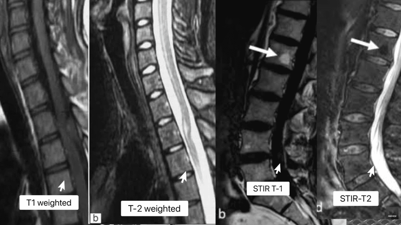
Our clinic’s protocol, based on expert sources like Apex Spine and Neurosurgery’s MVA treatment pages, includes T1-weighted, T2-weighted, STIR (Short Tau Inversion Recovery), and contrast-enhanced sequences to analyze complex findings.
- T1-Weighted Images: Optimal for anatomy, where fat appears bright and water/CSF dark. This sequence excels at identifying fatty degeneration, such as in Modic Type 2 changes or epidural lipomatosis, a condition associated with steroid use but exacerbated in MVAs. As per the MVA specialist page from Palm Harbor Ortho, T1 helps delineate contours and continuity with the epidural venous plexus.
- T2-Weighted Images: Ideal for pathology, with fluid, edema, and healthy disc nuclei appearing bright. This reveals heterogeneous signals in masses, making it crucial for distinguishing disc extrusions from venous dilations. Expert sites like OSI STL’s spine injury post-car accident guide note that T2 highlights annular tears as focal high-signal zones (HIZ), indicating water tracking through fissures—a direct sign of acute structural failure from MVA forces.
- STIR Images: Fat-suppressed T2 sequences that nullify fat signals, confirming bone marrow edema, inflammation, or soft tissue edema. NINDS emphasizes STIR’s sensitivity for detecting occult microfractures or ligamentous injuries, such as interspinous edema, which we use to prove recent trauma.
- Contrast-Enhanced MRI (T1 with Gadolinium) is invaluable for assessing vascular structures, as highly vascular tissues appear bright due to contrast enhancement. Mayo Clinic’s SCI treatment page says this is a good way to distinguish tumors, infections, or post-surgical scars from recurrent herniations, especially in MVA cases where there is suspicion of infection or metastasis via Batson’s plexus.
The step-by-step process: Upon referral, we order an MRI within days of the MVA to capture acute changes, as recommended by Las Vegas Spine & Pain Center’s MVA post. We look at the MRI images in a specific order: T1 to assess normal anatomy, T2 to detect any abnormalities, STIR to confirm swelling, and artifacts, such as chemical shifts at fat-water interfaces, are accounted for to avoid misdiagnosis.
For juries, we use annotated images: “See this bright spot on T2? That’s an annular tear from the crash force, like a tire blowout, causing nerve pain that keeps your client from work—impairment worth $X in lost wages.”
This protocol helps us find other problems, like a disc herniation that causes swelling in the veins, as mentioned on the New York Spine Specialist’s car accident page.
1.3 Key Complex MRI Findings We Interpret—Illuminating Hidden Impairments
Our expertise shines in interpreting nuanced findings, substantiated by diagnostic sources.
- Annular Tears and High-Intensity Zones (HIZ): Bright spots on T2 images in the back part of the disc show tears that let water from the – “High-intensity zone: a diagnostic sign of painful lumbar disc on magnetic resonance imaging.” This study validates HIZ as a marker of acute disruption, common in MVAs per AccidentDoctor.org.
- Modic Changes: Alterations in vertebral endplate marrow. Type 1 (edema): Dark on T1, bright on T2/STIR, indicating acute inflammation (0-8 weeks post-MVA). – “Modic Changes—From Inflammatory to Degenerative Processes.” Type 2 (fatty): Bright on T1/T2, dark on STIR (6-18 months). “Modic changes and their associations with clinical findings.”
- Interspinous Ligament Edema: Bright on STIR, from tensile forces in whiplash. “MRI assessment of whiplash injury.” Mayo Clinic’s whiplash page notes this as a pain generator.
- Facet Capsular Edema: High-intensity on T2/STIR around facets, specific for acute strain. – “Facet joint injuries in acute whiplash.”
- Spinal Cord Edema vs. Myelomalacia: Hyperintense on T2; edema (reversible contusion) vs. myelomalacia (irreversible necrosis with atrophy). – “MRI of spinal cord injury.” NINDS describes myelomalacia as leading to poor prognosis and permanent deficits.
- Multifidus Fatty Infiltration: Bright on T1, beginning 2-12 weeks post-injury due to denervation. – “Paraspinal muscle changes after injury.” Associated with chronic pain and instability.
- Vacuum Disc Phenomenon: Hypointense gas on all sequences, chronic marker. – “Vacuum phenomenon in degenerated discs.”
- Epidural Venous Plexus Pathology: Dilation appearing as fluid-like signals. PubMed link 9: “Batson’s plexus in spinal metastasis.” Differentiated from herniations using contrast.
- Spontaneous Regression of Herniated Discs: Extruded material becomes brighter on T2 due to inflammatory degradation. – “Spontaneous regression of intervertebral disc herniations.”
These interpretations reveal impairments like neurogenic claudication or sexual dysfunction, as per NINDS, translating to jury arguments for substantial damages.
Section 2: Providing Supporting Evidence for Causality and Injury Timing—Scientific Tools for Jury-Ready Narratives and Monetary Recoveries
Our clinic excels at furnishing Daubert-compliant evidence, using timelines to prove MVA causality and timing, as emphasized in expert witness directories such as Law.com’s spinal injury experts.
2.1 The Temporal Framework: Proximate Cause Through History and Onset
Causality begins with the temporal onset—symptoms appearing immediately after the MVA, consistent with Mayo Clinic’s whiplash symptoms. We document no prior complaints in the EMR, countering defense claims.
2.2 Modic Changes as Timestamps—Proving Recent Trauma
Modic Type 1 indicates acute endplate microfractures caused by MVA forces. Jury: “This edema is like a fresh bruise—it proves the accident caused the instability, leading to pain that impairs walking, warranting compensation for therapy and lost work.”
PubMed substantiation as above.
2.3 Wolff’s Law for Bone Remodeling—Debunking ‘Pre-Existing’ Claims
Wolff’s Law states that bone adapts to loads over 6+ months; osteophytes can’t form acutely. – “Wolff’s Law and bone’s structural adaptations.” Jury: “These spurs took months to grow—the new herniation beyond them is from the crash, causing pinching that disables your client.”
2.4 Acute-on-Chronic and Eggshell Skull – Aggravation Evidence
New extrusions beyond osteophytes prove aggravation, per OSI STL. The eggshell skull principle holds defendants liable for full damage.
Introduction: Turning MRI Images into Powerful Courtroom Evidence
When a client walks into your office after a motor vehicle accident (MVA), everything in their case eventually comes down to one core question:
Can you prove that this injury came from this crash, at this time, and that it truly explains your client’s pain, disability, and financial loss?
At Injury Medical Clinic PA in El Paso, my team and I have spent decades developing systems to answer that question with scientific precision. As a dual-licensed Chiropractor and Board-Certified Nurse Practitioner (DC, APRN, FNP-BC), my role is positioned at the intersection of
- Advanced spinal MRI interpretation
- Biomechanics and tissue-healing timelines
- Real-world functional impairments and disability
- The Daubert standard and medico-legal proof
Using a combination of high-level MRI analysis, Modic change interpretation, ligament and facet joint assessment, muscle and cord changes, and biomechanical timelines, we help attorneys in the El Paso community establish causality, date injuries, and defend medically necessary care.
The attached chapters on Advanced MRI Interpretation of Spinal Pathologies and Spinal Diagnostics: Causality, Injury Dating, MRI, and Biomechanics form the backbone of how we approach complex spinal cases, particularly after MVAs.
This article will walk you, as an attorney, through
- How MRI can distinguish acute traumatic injuries from old degeneration
- How we scientifically date injuries using bone, disc, ligament, and muscle changes
- How we present these findings in clear, jury-friendly language
- Why a dual-licensed NP/DC clinic in El Paso is uniquely positioned to support your cases and your clients’ monetary recoveries
- The Legal Foundation: MRI, Biomechanics, and the Daubert Standard
In personal injury litigation, you don’t just need a treating doctor—you need a spine expert who speaks both medicine and law.
Courts increasingly look for:
- Evidence grounded in peer-reviewed science, not anecdote
- Methods that are generally accepted in the medical and radiologic community
- A logical, testable cause-and-effect narrative between trauma and findings
That’s where advanced MRI interpretation comes in. MRI is considered the gold standard for evaluating soft tissues of the spine—discs, ligaments, nerve roots, spinal cord, and marrow. Studies show MRI has high accuracy (often 76–96%) for identifying disk herniations that correlate with surgical findings. (American Journal of Roentgenology)
From a legal standpoint, that means:
- We can show what was injured (disc, ligament, facet, cord, muscle).
- We can explain how it was injured (hyperflexion, hyperextension, compression, torsion). (ScienceDirect)
- We can estimate when those changes developed using established healing and remodeling timelines (e.g., Modic changes, bone spurs, muscle fatty infiltration). (PMC)
Our reports are built to satisfy both:
- Clinical standards—so patients receive appropriate, guideline-based care
- Medico-legal standards—so your evidence is defensible under Daubert or similar rules
- MRI 101 for Attorneys: Reading the Language of Spinal Imaging
You don’t need to become a radiologist—but understanding a few basics lets you quickly see why a case is strong.
2.1 Core MRI Sequences We Use
We routinely analyze:
- T1-weighted images
- Fat is bright
- Water/CSF is dark
- Great for anatomy and detecting fatty changes, marrow replacement, and chronic alterations
- T2-weighted images
- Fat and water are both bright
- Great for pathology: edema, fluid, acute disc hydration, and inflammation
- STIR/T2 fat-suppressed sequences
- Fat is suppressed (dark)
- Any bright signal that remains is water or edema—a key sign of acute or active inflammation (BioMed Central)
These sequences enable us to distinguish between:
- Acute injury (edema, inflammatory water, ligament tears, bone marrow edema)
- Chronic degeneration (fatty marrow, sclerosis, vacuum phenomenon)
Our internal protocols, as outlined in your attached documents, rely heavily on multi-sequence comparisons to avoid misreading normal anatomy, artifacts, or venous structures as herniations.
2.2 Batson’s Plexus and the Epidural Space: Why It Matters
The epidural venous plexus (Batson’s plexus) is a network of valveless veins in the spinal canal. Because these veins lack valves, blood can flow in either direction and act as a pathway for metastasis and infection. (NCBI)
For attorneys, this matters because:
- On MRI, engorged epidural veins (varices) can mimic a disc herniation.
- Without advanced interpretation, a radiology report might misidentify or under-describe the cause of canal narrowing or nerve root compression.
Our clinic is trained to differentiate:
- True disc extrusion vs.
- Venous engorgement vs.
- Epidural lipomatosis (excess fat)
by tracking how each structure behaves across T1, T2, and STIR sequences and, when appropriate, contrast-enhanced imaging.
- Establishing Causality: From Collision to Complaint to MRI
When we support an attorney, we don’t just say, “The MRI shows a herniated disc.” We build a causality narrative:
No prior symptoms → traumatic event → immediate or early onset of pain → objective MRI and exam findings that fit the biomechanics of that event.
3.1 Proximate Cause: Symptom Timeline and History
We document:
- No prior history of similar radiating pain, weakness, or numbness
- The exact mechanism of trauma (rear-end, T-bone, rollover, fall, work incident)
- The onset and progression of symptoms
- Emergency room and urgent care notes
- Past imaging (if available) for before/after comparison
As your attached diagnostics chapter emphasizes, simply asking, “Have you had this before?” isn’t enough. We proactively request prior records, including primary care and pediatric records if needed, to show a clean history before the collision.
3.2 Linking Imaging to the Mechanism
Different forces create different injury patterns:
- Hyperflexion/hyperextension whiplash
- Interspinous ligament edema
- Facet capsule sprain or effusion
- Possible annular tears and Modic changes in cervical vertebrae (ScienceDirect)
- Axial compression and flexion (typical in seat-belted lumbar trauma)
- Acute disc extrusion or sequestration
- Modic Type 1 changes in endplates
- Acute endplate injury and marrow edema (PMC)
- Translation/rotational injuries
- Discoligamentous injury: disruption of disc plus key stabilizing ligaments (Radiopaedia)
We tie the visible MRI pattern to a plausible and well-known mechanism of spinal trauma, which is exactly what jurors and judges need to see.
- MRI Signs That Help Us Date and Differentiate Injuries
This is where things become especially powerful for litigation. MRI doesn’t just say, “There is a problem.” It often tells us “how new” that problem is.
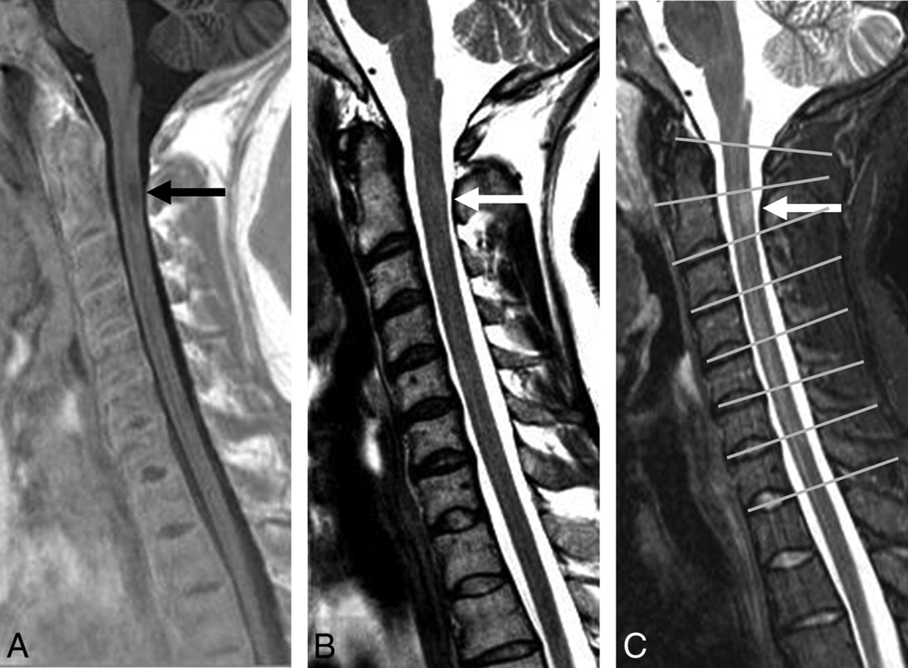
4.1 Annular Tears, High-Intensity Zones, and Acute Disc Injury
An annular tear appears as a bright focal signal (a “high-intensity zone”) in the normally dark outer ring of the disc on T2 or STIR images.
Research shows these high-intensity zones are strongly associated with painful discs and acute or subacute disc disruptions. (e-arm.org)
For attorneys, this means:
- If your client had no prior radicular symptoms and their post-collision MRI shows a herniation with a bright annular tear, we can support the argument that this is traumatic and recent, not just an “old degenerative bulge.”
Our own writings emphasize that a dark, “desiccated” disc may appear old, but if the extruded fragment is much brighter than the parent disc, it often indicates the water has been expelled into the herniation—consistent with a recent event.
4.2 Modic Changes: The Bone’s Timeline of Injury
Modic changes are signal alterations in the vertebral endplate and adjacent marrow that evolve over time:
- Type 1 (inflammatory/edematous)
- Dark on T1, bright on T2 / STIR
- Represents active inflammation and micro-instability (PMC)
- Usually appears in the early phase after disc/endplate injury
- Type 2 (fatty degeneration)
- Bright on T1 and T2, dark on STIR
- Reflects chronic changes and fatty replacement of marrow
- Typically develops over 6–18 months after insult (PMC)
- Type 3 (sclerotic)
- Dark on all sequences
- Indicates long-standing, heavily remodeled bone
Recent literature continues to confirm that these patterns correlate with vertebral endplate damage and vertebrogenic low back pain. (OUP Academic)
For medico-legal purposes:
- Modic 1 changes seen shortly after a crash support a recent traumatic event.
- Modic 2 changes on an MRI 12–18 months later can still be used to validate that the endplate was injured around the time of the accident, consistent with a long-term pain generator.
We use these timelines extensively when explaining to adjusters, defense experts, and ultimately juries.
4.3 Ligamentous Injuries: Interspinous & Facet Capsule Edema
Interspinous ligament injuries appear as bright signals between spinous processes on STIR or T2 fat-suppressed images. (BioMed Central)
Facet joint capsular injuries may show:
- Joint widening
- Bright fluid in the joint
- Surrounding edema (facet synovitis) (BioMed Central)
These are classic signs of acute or subacute trauma. They often correspond to:
- Local neck or back pain
- Pain with extension and rotation
- Referred pain patterns that jurors can easily relate to (“I can’t look over my shoulder anymore.”)
We highlight these injuries in our reports and, when testifying, use labeled images to visually show the inflamed ligaments or joints—so the jury can literally see the injury.
4.4 Spinal Cord Edema and Myelomalacia
In more severe cases—such as high-energy crashes or falls—the spinal cord itself may be bruised:
- Cord edema (contusion)
- Bright T2 signal inside the cord
- Represents active swelling and injury
- Myelomalacia
- Chronic aftermath: persistent T2 hyperintensity with cord thinning
- Associated with permanent neurological loss and poor prognosis
These findings are critical for explaining long-term disability, gait changes, hand function loss, or balance issues—key factors in future damages and life-care planning.
4.5 Muscle Atrophy and Fatty Infiltration
The paraspinal muscles—especially the multifidus—often undergo rapid atrophy and fatty infiltration after injury and disuse.
- On T1, fatty infiltration appears bright, replacing normal muscle.
- This can start as early as 2–12 weeks post-injury and is strongly associated with chronic instability and pain.
We use these changes to:
- Show that pain and disuse have led to structural muscle changes.
- Explain why some clients don’t “bounce back” despite conservative timelines pushed by insurers.
- Differentiating Acute Trauma from Old Degeneration
Defense arguments often hinge on one idea:
“Those MRI findings are old, degenerative changes that had nothing to do with this accident.”
We address this head-on by acknowledging degeneration honestly—then accurately distinguishing it from the new traumatic component.
5.1 “Acute-on-Chronic”: Using Osteophytes as Landmarks
Osteophytes (bone spurs) develop over many months in response to joint or disc instability, following Wolff’s law and piezoelectric principles.
Because it can take about six months for a spur to become radiographically visible, we use these structures as historical markers:
- If herniated disc material extends beyond the outer border of a mature osteophyte, that extension is almost certainly new.
- The spur shows the old “boundary”; the disc fragment beyond it is the fresh aggravation.
This acute-on-chronic pattern validates both:
- A pre-existing vulnerability, and
- A new, collision-related worsening that the defendant is still fully liable for under the “eggshell skull” principle.
5.2 Vacuum Disc Phenomenon vs. Acute Findings
A vacuum disc—gas inside the disc space—appears black on all sequences and is a sign of advanced degeneration, not acute trauma.
We often tell juries:
These black pockets resemble old cracks in a dried-out sponge—signs of long-standing wear and tear. But that doesn’t mean the recent tearing of this part of the disc (points to acute herniation) isn’t new or related to trauma.
By differentiating clearly between old and new, we enhance your credibility and defuse the “it’s all degenerative” narrative.
- Advanced MRI Concepts Attorneys Should Know (Without Becoming Radiologists)
To fully appreciate how deep we go for your cases, here are a few advanced points we routinely analyze.
6.1 Disc Extrusion & Biochemical Transformation
When a disc extrudes into the epidural space:
- Its proteoglycan-rich nucleus pulposus is exposed to immune cells, particularly mast cells.
- These cells break down proteoglycans, releasing free water and often making the extruded fragment brighter on T2 than the original disc.
Clinically, this means:
- A bright, hydrated extrusion soon after a crash strongly suggests a recent tear and displacement, not a long-standing, dried-out herniation.
6.2 Epidural Venous Plexus vs. “Mass”: Avoiding Misdiagnosis
The epidural venous plexus can enlarge or engorge:
- As a normal physiologic response to disc protrusion (dural “tenting” that creates negative pressure)
- As a true varix or in association with mass effect, sometimes mimicking a tumor or herniation on limited sequences (Frontiers)
We carefully examine:
- Continuity with known venous channels
- Enhancement patterns with contrast
- Shape and location relative to disc and dura
This level of detail allows us to refine or challenge radiology reads when necessary and make sure your case rests on the most accurate interpretation possible.
6.3 Chemical Shift Artifacts and Pitfalls
Not every bright or dark line indicates pathology. Chemical shift artifacts at fat–water interfaces, such as epidural fat and CSF, can resemble pathology if they are not recognized.
Our training and internal case-based analyses help us distinguish artifact from injury, so the opposing side can’t undermine your case by pointing out misread images.
- Case Types We Commonly Explain to Juries
Below are examples of case patterns we frequently see in El Paso personal injury litigation and how we present them.
7.1 Rear-End Whiplash with Cervical Facet & Ligament Injury
Scenario:
A 35-year-old office worker, rear-ended at a stoplight, develops neck pain, headaches, and shoulder radiation.
Key Findings:
- STIR images show interspinous ligament edema at C4–C6. (BioMed Central)
- Facet joints at C5–C6 demonstrate joint effusion and capsular edema, consistent with acute capsular sprain. (SpringerOpen)
- Early Modic 1 changes at the adjacent endplate (subtle marrow edema). (American Journal of Roentgenology)
How we explain it to a jury:
“In this image, the normally dark ligament between these bones has turned bright white—that’s water from inflammation, meaning the ligament was stretched or torn recently. These swollen facet joints are like sprained knuckles in the neck. These findings simply do not appear in people overnight without trauma.”
Why a dual NP/DC matters:
- As a chiropractor, I address mechanical alignment and soft-tissue rehabilitation.
- As a nurse practitioner, I coordinate imaging, prescribe medications when appropriate, manage headaches, and screen for concussion or neurological complications.
Attorneys use this combined expertise to justify extended treatment timelines, interventional pain management if needed, and more realistic non-economic damages.
7.2 Lumbar Disc Extrusion with Modic Changes After a T-Bone Collision
Scenario:
A 42-year-old warehouse worker is T-boned at an intersection. Soon afterward, he develops severe low back pain with left leg sciatica.
Key Findings:
- Large left paracentral L4–L5 disc extrusion compressing the L5 nerve root.
- Bright T2/STIR signal in the posterior annulus (annular tear). (e-arm.org)
- Modic 1 changes at the inferior L4 endplate. (PMC)
How we explain it:
“This bright mass here is disc material that has been forcefully pushed out of place and is crushing the nerve that feeds his leg. The bright streak at the back of the disc is the tear where that material escaped. The edema in the bone above is the body’s immediate reaction to that injury.”
We then relate his complaints—shooting pain, weakness, and numbness—to the exact dermatome and myotome served by the L5 nerve, connecting subjective symptoms to objective MRI.
Legal impact:
- Supports causality (no prior radicular pain, now clear nerve compression).
- Justifies surgery or higher-level interventions if needed.
- Substantiates lost earning capacity, especially for a physically demanding job.
7.3 “Minor” Impact, Major Injury in a Degenerative Spine (Eggshell Principle)
Scenario:
A 60-year-old with pre-existing degenerative changes is involved in a low-speed crash. Defense argues “pre-existing degeneration only.”
Key Findings:
- Pre-existing osteophytes and vacuum changes at L5–S1 (old degeneration).
- A new extrusion at L5–S1 that now extends beyond the prior osteophyte margin.
- Fresh Modic 1 changes and facet effusion at the same level.
We testify:
“These arthritic changes were there for years, but he was living and working with them. This accident didn’t create the degeneration—it pushed the already weakened tissue over the edge, creating a new disc herniation that now extends beyond the old bony overgrowth. The law doesn’t require a perfectly healthy spine to have a valid injury.”
This helps you:
- Reframe the defense argument.
- Show trauma superimposed on vulnerability, not a purely degenerative problem.
7.4 High-Energy Trauma with Spinal Cord Injury
Scenario:
A rollover crash leads to incomplete spinal cord injury.
Key Findings:
- Cord edema and, over time, myelomalacia at C5–C6.
- Multi-level ligamentous injury and potential discoligamentous instability (BioMed Central)
Here, our role expands into:
- Explaining permanent loss of function, muscle atrophy, and chronic pain.
- Connecting MRI changes to long-term impairment ratings and future medical needs.
- Providing expert testimony on life-care planning coordination with other specialists.
- Our Evaluation Process: How We Build a Medico-Legal Spine Case
Attorneys repeatedly return to our clinic because we do more than read scans—we build a narrative that is clinically sound and courtroom-ready.
8.1 Step 1: Comprehensive History and Record Retrieval
- Detailed mechanism of injury, seat position, headrest, and body position.
- Prior medical history, including old MRIs/X-rays if available.
- Medication use, prior pain management, prior motor vehicle accidents.
We use EMR integrations and direct record requests to demonstrate no prior similar complaints whenever possible.
8.2 Step 2: Dual-Scope Physical & Neurological Examination
As a DC + APRN, I combine:
- Orthopedic provocation tests
- Detailed neurological exam (strength, sensation, reflexes)
- Gait analysis, balance testing, functional assessments
- Identification of red flags requiring advanced imaging or specialty referral
This ensures we:
- Order targeted, medically necessary MRI studies, not generic panels.
- Identify the correct levels and structures to scrutinize.
8.3 Step 3: MRI Protocol, Review, and Second-Look Analysis
We work closely with imaging centers to ensure:
- Proper sequences (T1, T2, STIR/fat-sat, sometimes contrast)
- Appropriate slice thickness and planes
- Coverage of suspected injured regions
Then we:
- Review radiology reports and independently analyze the images.
- Cross-check for overlooked Modic changes, ligamentous injuries, subtle facet pathology, and epidural venous changes.
- Correlate imaging with exam and mechanism, using principles from the attached texts.
8.4 Step 4: Integrated Medical-Legal Reporting
Our reports are designed with attorneys and adjusters in mind:
- Plain-English explanation of each key MRI finding
- Clear statements on causality (“within a reasonable degree of medical probability …”)
- Discussion of injury dating, referencing known biological timelines
- Differentiation between pre-existing and new injuries
- Justification of treatment plans, including chiropractic care, medications, interventional procedures, rehab, and follow-up imaging
We often include annotated images that highlight:
- Herniations
- Ligament tears
- Modic changes
- Facet effusion
- Cord or nerve injuries
so that jurors and mediators can understand at a glance.
8.5 Step 5: Ongoing Care and Outcome Documentation
Over time, we:
- Track pain scores, functional measures, and neurologic changes.
- Document treatment response, flares, and plateaus.
- Re-image when clinically indicated to show progression, regression, or stability of findings. (AAFP)
This longitudinal data is invaluable when:
- Negotiating with adjusters
- Responding to defense IMEs
- Explaining permanent impairment and future care needs
- How Our Work Helps Attorneys Maximize Case Value
A well-documented MRI-based injury story does more than “win the argument.” It directly supports monetary recovery by:
- Demonstrating objective evidence that your client is injured
- Establishing clear causality tied to the crash date
- Defending reasonable, necessary treatment against denials
- Supporting future medical costs and life-care planning
- Anchoring pain and suffering in visible, structural changes
Because we operate within the El Paso community and the surrounding region, we understand:
- The local jury pool’s expectations and questions
- Common defense tactics used by regional insurers and IME providers
- How to present complex imaging in a way that is accurate, humble, and persuasive
- Why a Dual-Licensed NP & Chiropractor Clinic Is Ideal for PI Cases in El Paso
Attorneys increasingly seek out providers who bridge the gap between conservative musculoskeletal care and advanced medical diagnostics.
At Injury Medical Clinic PA:
- As a Chiropractor (DC), I understand the real-world biomechanics of the spine, kinetic chain, posture, and functional limitations.
- As a Board-Certified Nurse Practitioner (APRN, FNP-BC), I have the authority to:
- Order and interpret MRI, CT, and other advanced imaging
- Prescribe and manage medications
- Coordinate care with neurosurgeons, orthopedists, and pain specialists
- Address comorbidities such as diabetes, hypertension, and obesity that affect recovery
This dual role allows us to:
- Create integrated, evidence-based treatment plans.
- Provide coherent testimony that blends chiropractic, medical, and legal perspectives.
- Offer attorneys a single clinical voice who can speak across disciplines and explain the full story behind the imaging.
- Summary for Attorneys: What You Can Expect When You Collaborate with Us
When you refer your client to our clinic, our goals align with yours:
- Identify all trauma-related spinal injuries using advanced MRI and biomechanical analysis.
- Differentiate new injuries from old degeneration with scientifically grounded timelines (Modic changes, bone remodeling, and ligament and muscle changes).
- Explain findings in clear, jury-friendly language, supported by peer-reviewed literature. (PMC)
- Document a medically necessary, guideline-based treatment course, integrating chiropractic and medical care. (AAFP)
- Support you through negotiations, depositions, and trial, providing expert testimony that stands up under cross-examination.
Our mission is to make sure that:
- The full extent of your client’s disability and impairment is seen, understood, and believed.
- You have the scientific tools to connect that disability directly to the motor vehicle accident in question.
When juries can see the injury, hear the biomechanics, and understand the timeline, they are far more likely to appreciate the real human cost of spinal trauma—and to award damages that reflect that reality.
Selected Scientific & Clinical References (Hyperlinked)
Note: These are examples of the peer-reviewed and expert sources we routinely draw on to support injury dating and causality in our medico-legal work.
- Rahme R, Moussa R. Modic vertebral endplate and marrow changes: pathophysiology and clinical significance. Insights Imaging. (PMC)
- Crockett MT et al. Modic Type 1 changes and their clinical implications. AJR Am J Roentgenol. (American Journal of Roentgenology)
- Zhang YH et al. Modic changes: a systematic review. Eur Spine J. (PMC)
- Heggli I et al. Modic Type 1 changes and low back pain characteristics. J Orthop Translat. (ScienceDirect)
- Kumar Y et al. Role of MRI in acute spinal trauma. BMC Musculoskelet Disord. (BioMed Central)
- Benedetti PF et al. MR imaging findings in spinal ligamentous injury. AJR Am J Roentgenol. (American Journal of Roentgenology)
- Perolat R et al. Facet joint syndrome: imaging and interventional management. Insights Imaging. (SpringerOpen)
- Divi SN et al. MRI characteristics of lumbar disc herniation and symptom acuity. Int J Spine Surg. (PMC)
- Kim SY et al. Magnetic resonance findings of acute severe low back pain. Ann Rehabil Med. (e-arm.org)
- Lee J et al. Pain, disability, and MRI changes in lumbar disc herniation over time. Clin Orthop Surg. (PMC)
- Brook RC et al. Batson’s plexus and retrograde venous spread of malignancy. Clin Anat. (PMC)
- Bai J et al. Imaging of metastatic epidural spinal cord compression. Front Radiol. (Frontiers)
Post Disclaimer
General Disclaimer, Licenses and Board Certifications *
Professional Scope of Practice *
The information herein on "Advanced Spinal MRI Interpretation and Medico-Legal Expertise: Empowering Attorneys with Dr. Alex Jimenez's Expert Approach to Proving Causality, Timing, and True Impairment in Motor Vehicle Accident Injury Cases (2025 Comprehensive Guide)" is not intended to replace a one-on-one relationship with a qualified health care professional or licensed physician and is not medical advice. We encourage you to make healthcare decisions based on your research and partnership with a qualified healthcare professional.
Blog Information & Scope Discussions
Welcome to El Paso's Premier Wellness and Injury Care Clinic & Wellness Blog, where Dr. Alex Jimenez, DC, FNP-C, a Multi-State board-certified Family Practice Nurse Practitioner (FNP-BC) and Chiropractor (DC), presents insights on how our multidisciplinary team is dedicated to holistic healing and personalized care. Our practice aligns with evidence-based treatment protocols inspired by integrative medicine principles, similar to those on this site and on our family practice-based chiromed.com site, focusing on naturally restoring health for patients of all ages.
Our areas of multidisciplinary practice include Wellness & Nutrition, Chronic Pain, Personal Injury, Auto Accident Care, Work Injuries, Back Injury, Low Back Pain, Neck Pain, Migraine Headaches, Sports Injuries, Severe Sciatica, Scoliosis, Complex Herniated Discs, Fibromyalgia, Chronic Pain, Complex Injuries, Stress Management, Functional Medicine Treatments, and in-scope care protocols.
Our information scope is multidisciplinary, focusing on musculoskeletal and physical medicine; wellness; contributing etiological viscerosomatic disturbances within clinical presentations; associated somato-visceral reflex clinical dynamics; subluxation complexes; sensitive health issues; and functional medicine articles, topics, and discussions.
We provide and present clinical collaboration with specialists from various disciplines. Each specialist is governed by their professional scope of practice and licensure jurisdiction. We use functional health & wellness protocols to treat and support care for musculoskeletal injuries or disorders.
Our videos, posts, topics, and insights address clinical matters and issues that directly or indirectly relate to our clinical scope of practice.
Our office has made a reasonable effort to provide supportive citations and has identified relevant research studies that support our posts. We provide copies of supporting research studies upon request to regulatory boards and the public.
We understand that we cover matters that require an additional explanation of how they may assist in a particular care plan or treatment protocol; therefore, to discuss the subject matter above further, please feel free to ask Dr. Alex Jimenez, DC, APRN, FNP-BC, or contact us at 915-850-0900.
We are here to help you and your family.
Blessings
Dr. Alex Jimenez DC, MSACP, APRN, FNP-BC*, CCST, IFMCP, CFMP, ATN
email: [email protected]
Multidisciplinary Licensing & Board Certifications:
Licensed as a Doctor of Chiropractic (DC) in Texas & New Mexico*
Texas DC License #: TX5807, Verified: TX5807
New Mexico DC License #: NM-DC2182, Verified: NM-DC2182
Multi-State Advanced Practice Registered Nurse (APRN*) in Texas & Multi-States
Multi-state Compact APRN License by Endorsement (42 States)
Texas APRN License #: 1191402, Verified: 1191402 *
Florida APRN License #: 11043890, Verified: APRN11043890 *
Colorado License #: C-APN.0105610-C-NP, Verified: C-APN.0105610-C-NP
New York License #: N25929, Verified N25929
License Verification Link: Nursys License Verifier
* Prescriptive Authority Authorized
ANCC FNP-BC: Board Certified Nurse Practitioner*
Compact Status: Multi-State License: Authorized to Practice in 40 States*
Graduate with Honors: ICHS: MSN-FNP (Family Nurse Practitioner Program)
Degree Granted. Master's in Family Practice MSN Diploma (Cum Laude)
Dr. Alex Jimenez, DC, APRN, FNP-BC*, CFMP, IFMCP, ATN, CCST
(Board Certified: Family Practice Nurse Practitioner—Multistate)*
(Licensed Nurse Practitioner & Chiropractor - Multistate)*
Clinical Director
Digital Business Card
Dr. Maria Cardenas, MD
(Board Certified: Internal Medicine)
(Licensed Medical Doctor)
Medical Director, Clinical Director & Collaborative Physician
NPI # 1164426749
MD License #: J2933
Licenses and Board Certifications:
MD: Medical Doctor
DC: Doctor of Chiropractic
APRNP: Advanced Practice Registered Nurse
FNP-BC: Family Practice Specialization (Multi-State Board Certified)
RN: Registered Nurse (Multi-State Compact License)
CFMP: Certified Functional Medicine Provider
MSN-FNP: Master of Science in Family Practice Medicine
MSACP: Master of Science in Advanced Clinical Practice
IFMCP: Institute of Functional Medicine
CCST: Certified Chiropractic Spinal Trauma
ATN: Advanced Translational Neutrogenomics
Memberships & Associations:
TCA: Texas Chiropractic Association: Member ID: 104311
AANP: American Association of Nurse Practitioners: Member ID: 2198960
ANA: American Nurse Association: Member ID: 06458222 (District TX01)
TNA: Texas Nurse Association: Member ID: 06458222
NPI: 1205907805
| Primary Taxonomy | Selected Taxonomy | State | License Number |
|---|---|---|---|
| No | 111N00000X - Chiropractor | NM | DC2182 |
| Yes | 111N00000X - Chiropractor | TX | DC5807 |
| Yes | 363LF0000X - Nurse Practitioner - Family | TX | 1191402 |
| Yes | 363LF0000X - Nurse Practitioner - Family | FL | 11043890 |
| Yes | 363LF0000X - Nurse Practitioner - Family | CO | C-APN.0105610-C-NP |
| Yes | 363LF0000X - Nurse Practitioner - Family | NY | N25929 |
Dr. Alex Jimenez, DC, APRN, FNP-BC*, CFMP, IFMCP, ATN, CCST
(Board Certified: Family Practice Nurse Practitioner—Multistate)*
(Licensed Nurse Practitioner & Chiropractor - Multistate)*
Clinical Director
Digital Business Card
Dr. Maria Cardenas, MD
(Board Certified: Internal Medicine)*
(Licensed Medical Doctor)*
Medical Director, Clinical Director & Collaborative Physician
NPI # 1164426749
MD License #: J2933
📆 Schedule Appointment: Schedule 24/7 (Click Here)