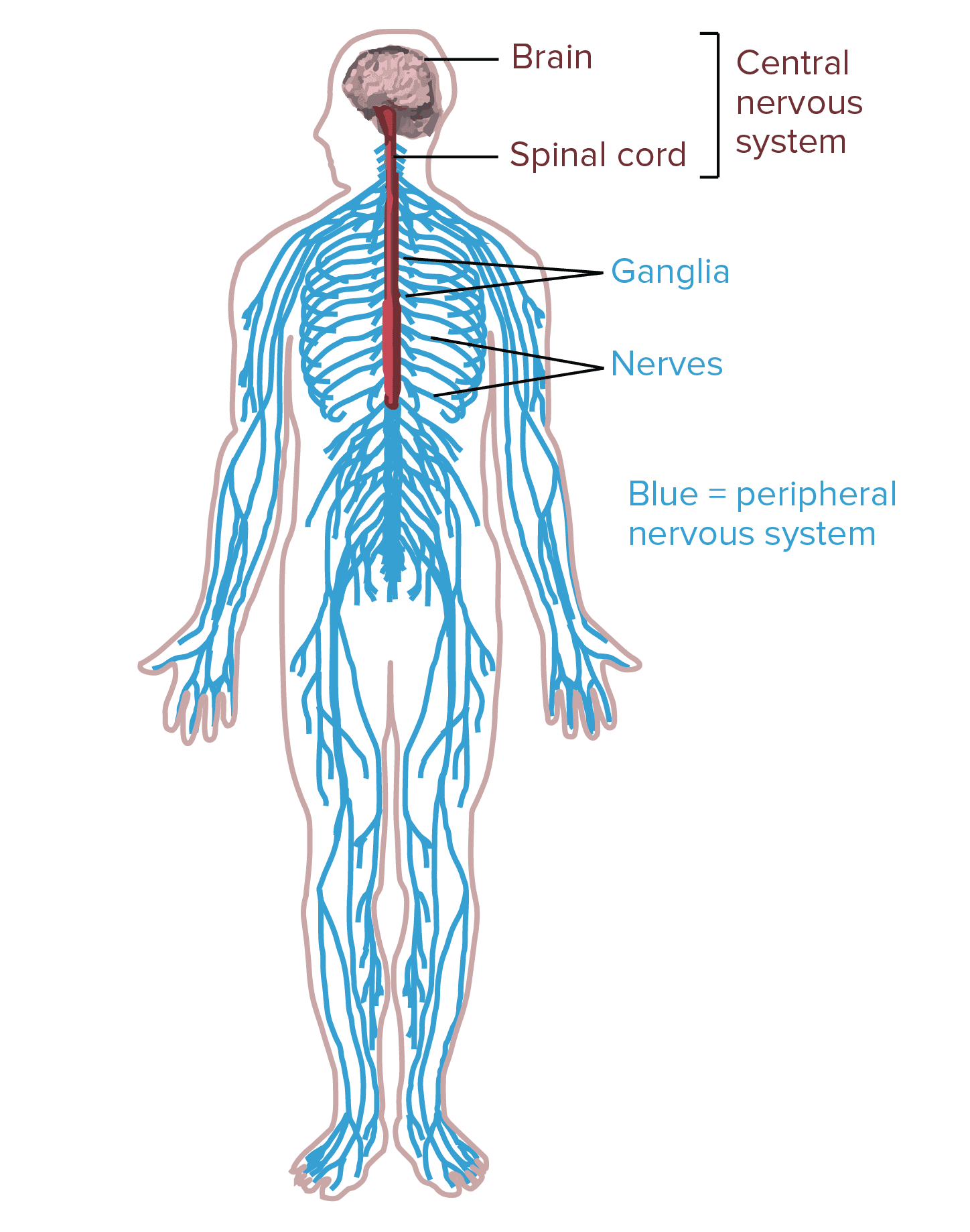
In humans, the nervous system consists of the central nervous system and the peripheral nervous system. The central nervous system, or CNS, consists of the brain and the spinal cord. It is in the CNS where the review of information occurs. The peripheral nervous system, or PNS, consists of the neurons and parts of neurons outside the CNS, including sensory neurons and motor neurons. Sensory neurons bring signals into the CNS, and motor neurons carry signals out of the CNS.
The cell bodies of PNS neurons, such as the motor neurons which control skeletal muscles, are found in the CNS. These motor neurons have long extensions, known as axons, which run from the CNS all the way to the muscles with which they connect with or innervate. The cell bodies of additional PNS neurons, such as the sensory neurons which provide information on touch, pain, position, and temperature, are found outside the CNS, in which they are found in clusters known as ganglia. The axons of peripheral nerves which run through a common pathway are bundled together to form nerves.

Table of Contents
Types of Neurons
According to their roles, the neurons within the human nervous system can be separated into three different categories, including the sensory neurons, the motor neurons, and the interneurons. Below, we will describe the types of neurons.
Sensory Neurons
The sensory neurons get information about what’s going on inside and outside the human body and they bring that information into the CNS where it could become processed. By way of instance, if you pick up a hot coal, the sensory neurons with nerve endings in your fingertips would communicate the information to your CNS that the hot coal is really hot.
Motor Neurons
The motor neurons get information from other neurons and they communicate commands to your muscles, organs, and glands. In the previous circumstance where you picked up a hot coal, the motor neurons innervating the structures on your fingers would cause your hand to let go of the hot coal. This is only one example of the role of motor neurons.
Interneurons
The interneurons, which can only be found in the CNS, connect one neuron to another. They get information from other neurons and communicate information to other neurons. When picking up a hot coal, the signals from the sensory neurons in your palms communicate to the interneurons on the spinal cord. Several of these interneurons communicate to the motor neurons controlling your finger muscles and cause your hand to let go of the hot coal. The motor neurons may communicate the signals to the interneurons in the spinal cord where it would ultimately create the perception of pain in the brain.
Interneurons are the most numerous types of neurons and they are involved in processing information, both through basic neural circuits, such as those triggered by picking up a hot coal, as well as in much more complicated circuits in the brain. Different combinations of interneurons in the brain and spinal cord allow you to draw the conclusion that objects which look similar to a lump of hot coal shouldn’t be picked up and they will also help keep that information for future reference.
Anatomy of a Neuron
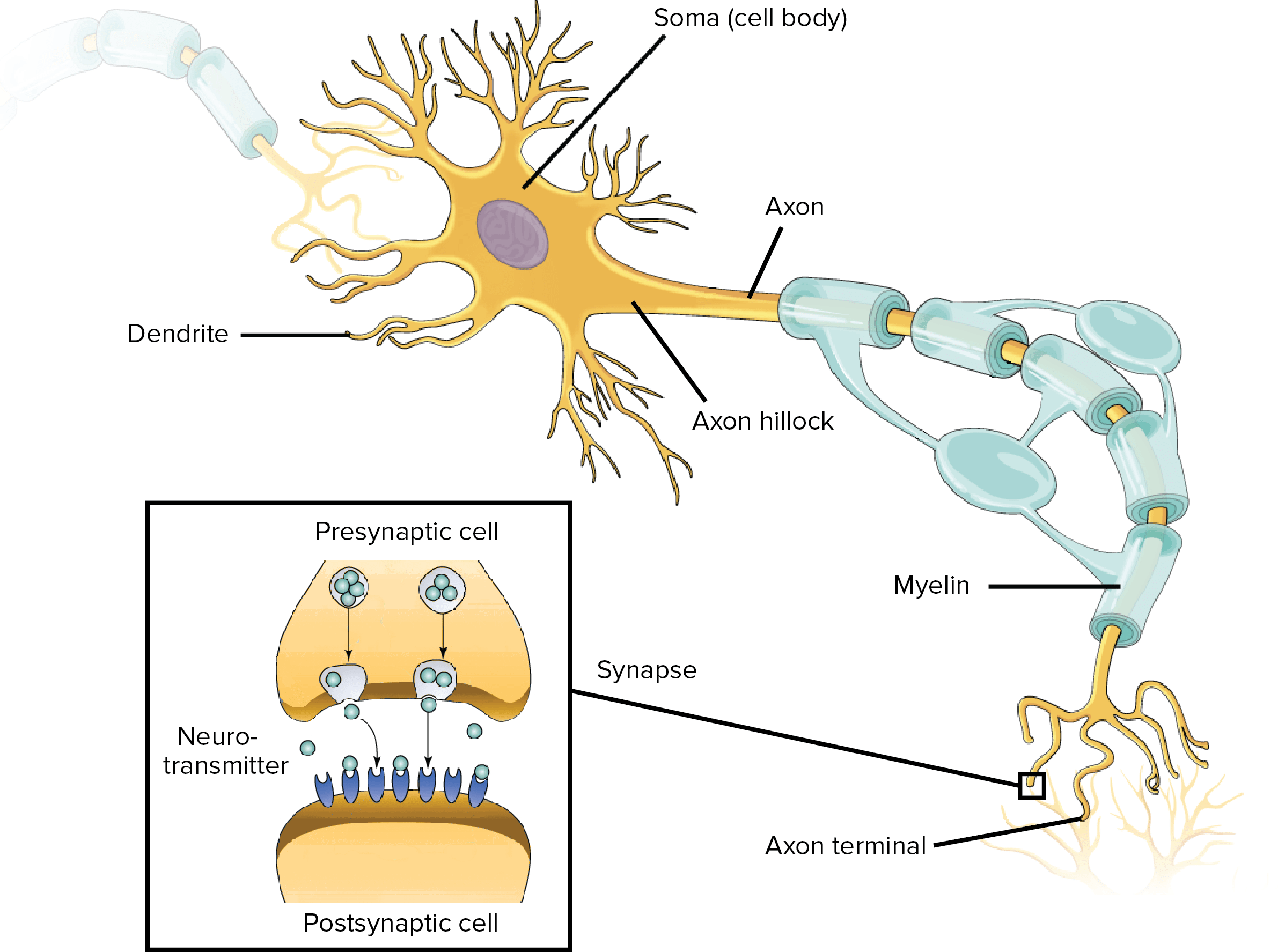
Neurons, similar to other cells, consist of a cell body known as the soma. The nucleus of the neuron is found in the soma. Neurons need to create proteins and most neuronal proteins are synthesized in the soma. Various processes, known as appendages or protrusions, run from the cell body. These include many small, branching processes, known as dendrites, and another process which is generally longer than the dendrites, known as the axon. It is possible to generalize that most neurons have three standard functions. These neuronal functions are mirrored in the anatomy of the neuron, including:
- Communicating information or signals.
- Combining incoming signals to determine whether or not the information should be passed along.
- Communicate information or signals to target cells, including muscles, glands, or other neurons.

Dendrites
The first two functions of the neuron, receive and process incoming signals or information, generally occur in the dendrites and cell body. Incoming signals can be either excitatory, which means that they tend to make the neuron generate an electrical impulse, or even inhibitory, which means that they tend to keep the neuron from generating an electrical impulse.
Most neurons receive many incoming signals or information throughout the dendrites. A single neuron can have more than one pair of dendrites and they may receive thousands of incoming information or signals. Whether or not a neuron is excited into firing an electrical impulse is dependent on the amount of each of the excitatory and inhibitory signals, or information, it receives. If the neuron does end up firing an electrical impulse, the action potential or nerve impulse runs down the axon.
Axons
The axon separates into many branches and develops bulbous swellings known as axon terminals or neural terminals. These axon terminals communicate with target cells. Axons are different from dendrites in several ways, as demonstrated below.
- The dendrites generally taper and are frequently covered with little bumps known as spines. The axon generally stays the same diameter for most of its length and doesn’t have spines.
- The axon exits from the cell body through a special region known as the axon hillock.
- Last but not least, many axons are covered with a special insulating compound known as the myelin, which helps them communicate the nerve impulse quickly. The myelin is never found on dendrites.
Synapses
Neuron-to-neuron communications are created on the dendrites and cell bodies of other neurons. These connections, known as synapses, are regions where information is taken from the first neuron, or the presynaptic neuron, to the target neuron, or the postsynaptic neuron. The synaptic connections between neurons and skeletal muscles are known as neuromuscular junctions and the connections between neurons and smooth muscle cells or glands are known as neuroeffector junctions.
Signals communicate through chemical messengers known as neurotransmitters. When an action potential runs down an axon and reaches the axon terminal, it triggers the release of neurotransmitters from the presynaptic cell. Neurotransmitters run through the synapse and connect to membrane receptors on the postsynaptic cell, communicating excitatory or inhibitory information. The first two basic functions of the neuron are important for the third basic function of the neuron.
The third function of the neuron, communicating signals to target cells, is also completed through the function of the axon and the axon terminals. Just as one neuron may communicate through many presynaptic neurons, it may also ultimately communicate through synaptic connections on numerous postsynaptic neurons throughout different axon terminals.

Glial Cells
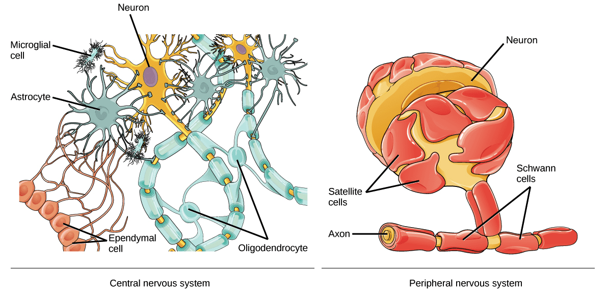
The glia, or glial cells, are fundamental to the nervous system. There are more glial cells in the brain than there are neurons. There are four types of glial cells in the adult human nervous system. Three of these, the astrocytes, the oligodendrocytes, and the microglia, are only found in the central nervous system or the CNS. The fourth, the Schwann cells, are only found in the peripheral nervous system or the PNS. Below, we will discuss the four types of glial cells, or glia, and their functions.
Astrocytes are the most numerous types of glial cell. There are also many different types of astrocytes and they each have a variety of different functions, such as regulating blood flow in the brain, maintaining the composition of the fluid which surrounds the neurons, and maintaining communications between nerves in the synapse. During development, astrocytes help neurons find their way and add to the development of the blood-brain barrier, which also helps protect the brain.
Microglia are associated to the macrophages of the immune system and act as scavengers to remove dead cells and debris.
The oligodendrocytes of the CNS and the Schwann cells of the PNS share a similar function. Both types of glia, or glial cells, create myelin, or the insulating compound which develops a sheath around the axons of many neurons. Myelin increases the speed with which an action potential runs down the axon and it plays a fundamental role in nervous system function.
Additional types of glial cells, along with the four main types of glia, include satellite glial cells and ependymal cells.
Satellite glial cells cover the cell bodies of neurons in PNS ganglia. Satellite glial cells are believed to support the role of the nerves and function as a protective barrier, however, their role is still misunderstood. Ependymal cells, which line the ventricles of the brain and the central canal of the spinal cord, have hairlike cilia which help improve the flow of the cerebrospinal fluid found within the ventricles and spinal tract. The human nervous system is necessary for our function.

Neurons are special cells found within the nervous system which communicate with other neurons in unique ways. The neuron is the basic working unit of the brain and it is designed to communicate information, or signals, to muscles, organs, gland, and other nerve cells. Most neurons consist of a cell body, an axon, and dendrites. The cell body contains the nucleus and the cytoplasm. Understanding the structure and function of the neuron is fundamental for overall health and wellness. – Dr. Alex Jimenez D.C., C.C.S.T. Insight
Diet and Exercise for Neurological Disease
The purpose of the article above is to discuss the purpose of functional neurology in the treatment of neurological disease. Neurological diseases are associated with the brain, the spine, and the nerves. The scope of our information is limited to chiropractic, musculoskeletal and nervous health issues as well as functional medicine articles, topics, and discussions. To further discuss the subject matter above, please feel free to ask Dr. Alex Jimenez or contact us at 915-850-0900 .
Curated by Dr. Alex Jimenez
Additional Topic Discussion: Chronic Pain
Sudden pain is a natural response of the nervous system which helps to demonstrate possible injury. By way of instance, pain signals travel from an injured region through the nerves and spinal cord to the brain. Pain is generally less severe as the injury heals, however, chronic pain is different than the average type of pain. With chronic pain, the human body will continue sending pain signals to the brain, regardless if the injury has healed. Chronic pain can last for several weeks to even several years. Chronic pain can tremendously affect a patient’s mobility and it can reduce flexibility, strength, and endurance.
Formulas for Methylation Support
XYMOGEN’s Exclusive Professional Formulas are available through select licensed health care professionals. The internet sale and discounting of XYMOGEN formulas are strictly prohibited.
Proudly, Dr. Alexander Jimenez makes XYMOGEN formulas available only to patients under our care.
Please call our office in order for us to assign a doctor consultation for immediate access.
If you are a patient of Injury Medical & Chiropractic Clinic, you may inquire about XYMOGEN by calling 915-850-0900.
For your convenience and review of the XYMOGEN products please review the following link.*XYMOGEN-Catalog-Download
* All of the above XYMOGEN policies remain strictly in force.
Post Disclaimer
Professional Scope of Practice *
The information on this blog site is not intended to replace a one-on-one relationship with a qualified healthcare professional or licensed physician and is not medical advice. We encourage you to make healthcare decisions based on your research and partnership with a qualified healthcare professional.
Blog Information & Scope Discussions
Welcome to El Paso's Premier Wellness and Injury Care Clinic & Wellness Blog, where Dr. Alex Jimenez, DC, FNP-C, a board-certified Family Practice Nurse Practitioner (FNP-BC) and Chiropractor (DC), presents insights on how our team is dedicated to holistic healing and personalized care. Our practice aligns with evidence-based treatment protocols inspired by integrative medicine principles, similar to those found on this site and our family practice-based chiromed.com site, focusing on restoring health naturally for patients of all ages.
Our areas of chiropractic practice include Wellness & Nutrition, Chronic Pain, Personal Injury, Auto Accident Care, Work Injuries, Back Injury, Low Back Pain, Neck Pain, Migraine Headaches, Sports Injuries, Severe Sciatica, Scoliosis, Complex Herniated Discs, Fibromyalgia, Chronic Pain, Complex Injuries, Stress Management, Functional Medicine Treatments, and in-scope care protocols.
Our information scope is limited to chiropractic, musculoskeletal, physical medicine, wellness, contributing etiological viscerosomatic disturbances within clinical presentations, associated somato-visceral reflex clinical dynamics, subluxation complexes, sensitive health issues, and functional medicine articles, topics, and discussions.
We provide and present clinical collaboration with specialists from various disciplines. Each specialist is governed by their professional scope of practice and their jurisdiction of licensure. We use functional health & wellness protocols to treat and support care for the injuries or disorders of the musculoskeletal system.
Our videos, posts, topics, subjects, and insights cover clinical matters and issues that relate to and directly or indirectly support our clinical scope of practice.*
Our office has made a reasonable effort to provide supportive citations and has identified relevant research studies that support our posts. We provide copies of supporting research studies available to regulatory boards and the public upon request.
We understand that we cover matters that require an additional explanation of how they may assist in a particular care plan or treatment protocol; therefore, to discuss the subject matter above further, please feel free to ask Dr. Alex Jimenez, DC, APRN, FNP-BC, or contact us at 915-850-0900.
We are here to help you and your family.
Blessings
Dr. Alex Jimenez DC, MSACP, APRN, FNP-BC*, CCST, IFMCP, CFMP, ATN
email: coach@elpasofunctionalmedicine.com
Licensed as a Doctor of Chiropractic (DC) in Texas & New Mexico*
Texas DC License # TX5807
New Mexico DC License # NM-DC2182
Licensed as a Registered Nurse (RN*) in Texas & Multistate
Texas RN License # 1191402
ANCC FNP-BC: Board Certified Nurse Practitioner*
Compact Status: Multi-State License: Authorized to Practice in 40 States*
Graduate with Honors: ICHS: MSN-FNP (Family Nurse Practitioner Program)
Degree Granted. Master's in Family Practice MSN Diploma (Cum Laude)
Dr. Alex Jimenez, DC, APRN, FNP-BC*, CFMP, IFMCP, ATN, CCST
My Digital Business Card


