
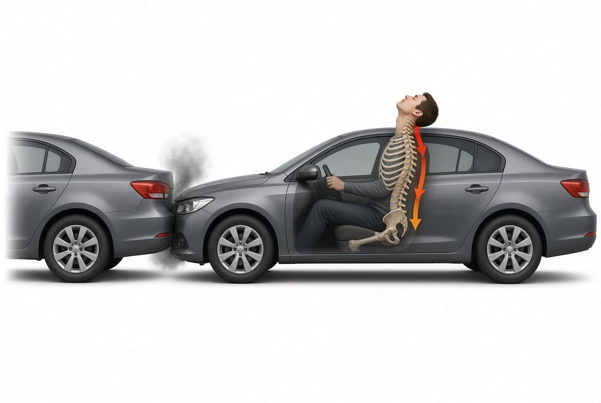
How one provider who holds two licenses, as a doctor of chiropractic and as a board-certified family nurse practitioner, worked with a multidisciplinary team to guide a young driver from an 8 out of 10 back injury to a stable, low-pain recovery, and why the care plan still accounts for the residual problems that remained.
Mechanism: Stopped car struck from behind by a larger SUV; driver restrained and facing forward, head restraint set low
Time to first visit: 11 days after the collision, after a brief visit to another clinic
Course of care: 12 visits over about 10 weeks, twice weekly at first and then weekly
Pain at intake: 4 out of 10 overall at the first visit; 8 out of 10 in the back three days later
Last rating: 1 out of 10 in the mid and low back; no neck pain
Outcome: Stable and released to self-management, with residual lumbar joint irritation and flare-ups under load
Most people picture a whiplash injury as a sore neck that fades in a week or two. Sometimes that is exactly what happens. Often, though, a rear-impact collision loads the whole spine at once: the neck, the upper back between the shoulder blades, the mid back and the low back, along with the muscles that tie the shoulders to the spine. The injury that hurts most at the start is not always the one that lingers. In this case, the neck settled quickly. The mid and low back did not settle as completely, and the patient’s final examination showed why.
This article walks through that case from the moment of impact to the final examination. It explains what was injured, how the team decided what to do and in what order, what each treatment was meant to accomplish, what the research says about each, and how the patient’s pain and function changed week by week. It is written for patients and families who want to understand this kind of injury, and for anyone curious about how an integrated practice that blends chiropractic and medical care approaches it. The patient’s identity has been removed, as described at the end of the page.
Why this case is worth telling
This is not a dramatic case. There was no fracture, no surgery, and no hospital stay. That is exactly why it is useful. Most collision injuries look like this one: a person who walks away from the scene, feels sore the next day, worsens over the following week, and then improves with good care. What happens in the weeks that follow decides whether the injury fades completely or leaves a back that flares every time a heavy box needs moving. This case shows the decisions that shape that outcome: how quickly care starts, how carefully the patient is screened, how the plan changes as pain falls, whether the factors outside the injured joint are addressed, and whether the final examination looks for what remains or confirms that resting pain is low.
It also shows something patients often find reassuring and frustrating at the same time. Pain can fall quickly, sometimes dramatically, while the underlying tissue is still healing and still sensitive under load. Understanding the gap between how an injury feels at rest and how it behaves under load is one of the most useful takeaways from a case like this.
Who treated the patient?
The patient was treated by Dr. Alexander Jimenez, DC, APRN, FNP-BC, who practices with two separate licenses. As a doctor of chiropractic, he examines and treats the spine, joints, and soft tissues with manual care, rehabilitation, and lifestyle guidance. As a board-certified family nurse practitio,ner he can take a medical history the way a primary care clinician does, screen for conditions that do not belong in a chiropractic office, review and manage medications, order and interpret laboratory and imaging studies, and make referrals to medical specialists. Having both skill sets in one person matters most in the first weeks after a crash, when the question is not only “what hurts” but “what else could be going on.”
He does not work alone. The practice is multidisciplinary. It includes medical providers, among them a board-certified internal medicine physician, and it offers the medical services of a family practice and internal medicine office alongside chiropractic care, rehabilitation, nutrition, naturopathic medicine and acupuncture. Patients can see a nurse practitioner and a chiropractor under one roof, and the team shares one record, so the person who adjusts the spine and the person who reviews the medications are reading the same notes.
Holistic and allopathic, not one or the other
Our practice aligns itself with both a holistic model and an allopathic (conventional medical) model, and we see ourselves as different from offices that practice only one. A purely holistic office may miss a medical problem that needs a prescription, a scan or a specialist. A purely conventional office may hand a patient a muscle relaxant and a note to rest, without the hands-on care, movement retraining and whole-person attention that often decide whether a neck or back injury heals cleanly. We would rather do both. In practice that meant, for this patient, that the same visit could include a medical screen for warning signs, a review of the medications another clinic had prescribed, a spinal examination with orthopedic and neurological testing, manual treatment, and advice about sleep, sitting, body weight and stress.
This is not a slogan. It shows up in specific decisions, and the rest of this case points them out: screening for concussion after the head struck the seat, deciding when imaging was and was not needed, deciding when to step treatment down, measuring body composition and foot mechanics because they change how an injured spine is loaded, and acting on new findings at the final visit rather than simply closing the file.
Why integration matters after a crash
Car-crash injuries sit at the border between musculoskeletal care and medicine. The pain is usually mechanical: sprained ligaments, irritated spinal joints, strained and guarding muscles. But the first job is to make sure nothing more serious is hiding behind it. A head strike raises the question of concussion. Numbness raises the question of nerve-root injury. Chest or abdominal pain after a belt load raises questions that are not chiropractic at all. Medications prescribed elsewhere need to be reviewed for interactions and side effects. A provider trained in both fields, working in a team with physicians, can answer those questions in the same office and the same week, rather than sending the patient on a series of separate appointments that may never be connected.
Integration also matters later, when recovery stalls or a new finding appears. An integrated team can change course quickly: add a referral, order a study, adjust medication or shift the treatment emphasis, without starting over with a new provider who has to learn the case from scratch.
The integrated team’s roles in this case
| Role | What that role contributed |
|---|---|
| Dual-licensed provider (DC and FNP-BC) | Medical and chiropractic history, red-flag screening, concussion screening, medication review, orthopedic and neurological examinations, spinal manipulation, care planning, final evaluation and recommendations |
| Chiropractic and rehabilitation staff | Soft-tissue therapy, therapeutic massage, mechanical traction, cold therapy, electrical stimulation, muscle-energy and stretching techniques, home exercise instruction |
| Medical team | Available for medical evaluation, medication management and referral when a finding calls for it; shares the same record |
| Assessment technology | Body composition analysis at the start and end of care; computerized foot-pressure and posture scanning |
| Outside specialists | Imaging and a pain-management consultation were arranged outside the practice; results and recommendations were folded into the plan |
How integrated care compares with the usual path
| Step | A typical fragmented path | The integrated path in this case |
|---|---|---|
| First evaluation | Urgent care or emergency visit, prescription, advice to rest | Full history and examination by one provider trained in both medicine and chiropractic |
| Screening for serious injury | Often limited to the first visit | Repeated at each stage: head injury, nerve signs, new symptoms |
| Medication | Prescribed by one office, rarely revisited | Reviewed alongside the treatment plan; non-drug options emphasized |
| Hands-on care | Separate referral, separate records | Same office, same record, same day |
| Whole-person factors | Seldom measured | Body composition, foot mechanics, posture, sitting habits and stress addressed |
| When recovery stalls | New referral, start over | Plan adjusted by the team that already knows the case |
| At discharge | File closed | Final orthopedic and neurological exam, residuals documented, clear next steps |
None of this guarantees a particular outcome. Injuries heal at different speeds in different people, and some leave lasting effects no matter how well they are treated. What integration offers is fewer blind spots and faster course corrections.
The collision and why the mechanism injures
The patient, a man in his late twenties, was the restrained driver of a small passenger car. His car was stopped when a mid-size sport utility vehicle struck it from behind at an estimated 15 to 25 miles per hour. The airbag did not deploy. He was looking straight ahead and was not braced for the impact. He recalled that the head restraint was set low relative to the back of his head, and his head and neck struck the seat and head restraint. He did not lose consciousness. Both vehicles had slight visible damage, and his car was driven from the scene.
At the time of the collision he felt discomfort at the back of the head, in the neck, across the back of both shoulders, in the middle of the back and in the middle of the low back. He also described soreness, shock and disbelief. Like many people in this situation, he did not feel severe pain at the scene; the pain and stiffness built over the following day and days.
What happens to the body in a rear impact
In a rear impact, the struck car is pushed forward abruptly. The seat goes with it, and the seatback drives the occupant’s torso forward. The head, which sits on top of the neck like a heavy ball on a flexible stalk, lags behind for a fraction of a second. During that fraction of a second the neck is forced into an S-shaped curve, with the lower neck bending backward while the upper neck bends forward, and then into full backward bending (extension) before the head is thrown forward again into flexion. Laboratory and clinical research has identified the small facet joints at the back of each spinal segment, the spinal ligaments, the discs and the neck muscles as the structures injured by this motion.2
The neck is not the only region loaded. As the torso is compressed into the seatback and then rebounds against the lap-and-shoulder belt, the thoracic spine (upper and mid back) and the lumbar spine (low back) are bent, compressed and sheared as well. The muscles that span the shoulder blades and connect the shoulders to the neck, especially the trapezius, contract hard and fast to protect the spine and are often strained in the process. That explains why this patient’s earliest complaints mapped so closely onto the neck, both shoulder girdles, the mid back and the low back.
What the seat and belt do
Seatbelts save lives, and this patient was wearing his. In a rear impact, though, the belt plays a different role than in a frontal crash. As the seat pushes the torso forward, the body first sinks into the seatback, compressing the spine and ramping slightly upward. Then, as the seatback springs back, the torso rebounds forward into the lap-and-shoulder belt. The shoulder belt crosses one shoulder and the chest diagonally, so the rebound loads the two sides of the body unevenly, which helps explain why the muscles around both shoulders and across the upper back are so often involved. The lap belt anchors the pelvis while the upper body moves, which concentrates bending and shear forces in the lower back. None of this is a reason not to wear a belt; it simply explains why pain after a rear impact so often spreads beyond the neck.
Why head position and the head restraint matter
Two details of this crash increased the load on his neck. First, he was looking straight ahead and was not braced. People who see a crash coming often tense their muscles, which can change how forces travel through the spine; an unbraced occupant takes the full, unprepared acceleration. Second, his head restraint was set low. A head restraint works by catching the head early, before the neck has bent far backward. When it is too low or too far back, the head travels further before it is supported, and the neck bends further. Large insurance-claim studies have shown that better head-restraint position and seat design meaningfully reduce neck-injury claims in rear-end crashes.4
Why slight vehicle damage does not rule out injury
It is natural to assume that little visible damage means little force. Modern bumpers, however, are designed to absorb low-speed impacts without deforming, so external damage is a poor measure of the acceleration that reaches the people inside. Controlled studies with volunteers have shown that rear impacts with very small changes in speed, as low as 4 to 8 kilometers per hour, produced whiplash-associated symptoms in roughly a third of participants, with measurable clinical findings in both men and women.1 Real-world data from crash-pulse recorders likewise show that the peak acceleration of the struck car, rather than the visible damage, is what tracks with lasting neck injury.3 This patient’s car was stopped and lighter than the vehicle that hit it, and the estimated closing speed was well above the speeds in those volunteer studies. The pattern of his symptoms fits the mechanism.
Why the pain came on over the next day
Delayed onset is common and expected. Immediately after a collision, adrenaline and the body’s stress response suppress pain. Over the next 24 to 72 hours, inflammation develops in the injured ligaments and joints, the muscles around them tighten into protective guarding, and stiffness builds. In the volunteer studies mentioned above, participants were examined again a day after the impact precisely because symptoms often appear or worsen after the event.1 This patient was seen at another clinic within a few days, where he was examined and started on prescription anti-inflammatory and muscle-relaxant medication, and he began care in our office 11 days after the collision. His symptoms had been continuous from the day of the crash.
For more on how crash forces translate into specific injuries, see our articles on collision injury dynamics and whiplash injuries.
Understanding the injury
To understand why this patient’s neck recovered faster than his back, and why his final examination still found residual problems, it helps to understand what the spine is made of, which parts are injured in a collision, and how soft tissue heals.
The anatomy in plain terms
The spine is a stack of 24 movable bones (vertebrae) sitting on the sacrum at the base. There are seven in the neck (cervical spine), twelve in the upper and mid back (thoracic spine), each carrying a pair of ribs, and five in the low back (lumbar spine). Between most vertebrae is a disc, a tough fibrous ring with a gel-like center that absorbs load and allows movement. At the back of each segment are two small facet joints, one on each side, lined with cartilage and wrapped in a joint capsule. These joints guide and limit motion, and they carry a share of the load whenever the spine bends backward or twists.
Ligaments run along the front and back of the vertebrae and between the bony arches, holding the stack together. Muscles in layers, from short deep muscles that span one or two segments to long muscles that run the length of the spine, move and stabilize it. The trapezius, a broad muscle shaped like a kite, runs from the base of the skull and the neck down the mid back and out to each shoulder blade; it is one of the muscles most often strained in a rear impact. Spinal nerves leave the spine between each pair of vertebrae and travel to the arms, chest, abdomen and legs, carrying both sensation and the signals that drive muscles.
What is injured in a rear impact
- Ligaments (sprain). The ligaments of the neck, mid back and low back can be stretched beyond their normal range, producing small tears and inflammation. A sprain is an injury to a ligament.
- Facet joints and their capsules. The facet joints are compressed and pinched during the S-shaped motion of the neck and as the trunk rebounds. Their capsules are richly supplied with pain nerves, and facet joint injury is one of the best-documented sources of persisting pain after whiplash.2
- Muscles and tendons (strain). Muscles that contract suddenly to protect the spine can be strained. Even when they are not torn, they often stay in protective spasm for weeks.
- Discs. Discs can be strained or, less commonly, torn, particularly with combined bending and compression.
- Nerves. Nerve roots and the small ganglia next to them can be irritated, producing pain, numbness or tingling that travels into a limb. This patient reported some numbness in the early weeks, which resolved; by the final examination his nerve testing was normal.
How whiplash injuries are graded
The Quebec Task Force, a landmark international review, classified whiplash-associated disorders into grades that are still used today.5 Grade I is neck complaint with pain, stiffness or tenderness only. Grade II adds musculoskeletal signs found on examination, such as reduced range of motion and point tenderness. Grade III adds neurological signs, such as reduced reflexes, weakness or sensory loss. Grade IV involves fracture or dislocation. This patient’s neck injury fit grade II: he had pain and stiffness with reduced, painful motion and muscle spasm, without neurological signs. The same logic applies to his mid and low back, which showed painful, restricted motion, muscle spasm and, at the end of care, positive joint-loading tests, again without signs of nerve-root injury.
The phases of soft-tissue healing
Ligaments, joint capsules and muscles heal in overlapping phases, and good treatment changes to match each one.
Inflammatory phase
The first several days. Blood flow and swelling increase, the injured tissue is cleaned up, and pain and muscle guarding are at their highest. The goals are to calm pain and spasm, protect the tissue and keep gentle movement going.
Repair phase
Roughly the first two to six weeks. The body lays down new collagen to bridge the injured tissue. The new tissue is weak and disorganized at first. The goals are to restore motion and gradually reintroduce load so the new tissue forms along useful lines.
Remodeling phase
From several weeks to many months. Collagen is reorganized and strengthened in response to the loads placed on it. The goals are strength, endurance and confidence in movement, with the patient increasingly managing on his own.
Healed ligament tissue is rarely as strong or as elastic as the original, and a joint whose capsule has been injured can stay sensitive to the movements that hurt it in the first place. That is one reason residual symptoms after a whiplash injury are common, and why the remodeling phase matters so much: the tissue becomes as resilient as the loads it is trained to handle.
Sensitization: when the alarm system stays turned up
Pain is not a simple readout of tissue damage. After an injury, the nerves in and around the injured tissue become more sensitive, and the spinal cord and brain can turn up the volume on signals from that area. In most people this sensitization fades as the tissue heals. In some, it persists. Research on people recovering from whiplash has found that local mechanical tenderness over the injured neck segments is present early in nearly everyone, but in those who go on to recover slowly or incompletely, the sensitivity persists and can spread.9 This is part of why early, active, well-paced care and reassurance matter: they help the nervous system settle rather than stay on high alert.
Headache, dizziness and the head strike
This patient’s head struck the seat and head restraint, and he reported discomfort at the back of the head at the time of the collision. Any head strike raises the question of concussion, a mild traumatic brain injury that can cause headache, dizziness, nausea, difficulty concentrating, sleep disturbance and sensitivity to light or noise. Neck injuries can also cause headache, because the upper neck joints and muscles refer pain to the back of the head. He was screened for these symptoms at the start of care; he denied headache, nausea, dizziness and vomiting, and his head symptoms did not return during care. The screening is described in more detail later in this case.
Why the mid back and low back behave differently from the neck
The three regions of the spine are built for different jobs, and they respond to injury differently. The neck is built for mobility: it carries a head that weighs ten to twelve pounds and swivels through a large range of motion many thousands of times a day. Its muscles are relatively small, and its joints are oriented to allow rotation and tilting. After a sprain, the neck often recovers its comfort quickly once spasm settles and motion returns, because ordinary daily movement keeps loading it in small, varied doses, which is exactly what healing tissue needs.
The thoracic spine is built for stability. Its vertebrae are tied to the ribs, which form a cage around the heart and lungs. That cage limits motion, which protects the organs inside but also means that when the mid back stiffens after an injury, it tends to stay stiff unless it is deliberately mobilized. The muscles between the shoulder blades and along the mid back are also the ones most affected by long hours of sitting, especially with the head held forward, because they work constantly to hold the upper body upright.
The lumbar spine is built for load. It carries the weight of everything above it and transmits the forces of lifting, bending and carrying to the pelvis and legs. Its facet joints are oriented to resist twisting and to carry a share of the load when the spine bends backward. Those same joints are loaded by lifting, by standing for long periods and, in a different way, by sustained sitting. When a lumbar facet joint capsule has been sprained, it can become a pain source that is quiet at rest and flares under exactly those loads. That is the pattern this patient showed: low resting pain, with soreness after lifting boxes or moving furniture and ache after long periods of sitting.
In other words, the region that hurt most at the start, the back, was also the region whose job made it hardest to rest. The neck could move freely through the day and recover; the low back had to keep carrying his body weight, every chair he sat in and every box he lifted.
Muscles that guard and muscles that tire
After an injury, the muscles around the injured joints tighten automatically. This guarding is protective in the first days: it limits movement while the tissue is most vulnerable. If it persists, though, it becomes part of the problem. Persistently tight muscles fatigue, develop tender points, restrict circulation and pull on the joints they surround. The large muscles at the back of the thighs and calves can tighten as well, especially in someone who sits for long periods, and tight hamstrings change the way the pelvis tilts and the low back moves when bending. In this case, the examination in the fourth week found marked tightness in the low back, hips and legs, and the final examination still described the leg muscles as very tight. Treating that tightness, with soft-tissue therapy, muscle-energy techniques and stretching, is part of treating the low back.
Factors specific to this patient
Every injury happens to a particular body, and several features of this patient’s body and daily life were relevant to how his spine would heal.
- Body weight and composition. His body mass index at the start of care was about 30. Higher body weight increases the load on the lumbar spine with every step and every hour of sitting, and research has found that obesity is associated with more low back pain, more care-seeking for it, and more chronic low back pain.17 Body composition was measured at the start and end of care, as described below.
- Foot mechanics. A computerized foot-pressure scan showed that his arches collapsed more than optimal under load (moderate overpronation), with a difference in arch height between the two feet. The way the feet absorb load changes how forces reach the knees, hips, pelvis and lumbar spine.
- Posture. A posture scan showed a forward-head posture in the moderate range. A head held forward of the shoulders increases the load the neck and upper back muscles must carry.
- Prolonged sitting. His work involved long hours seated. Sitting for long periods places sustained load on the lumbar discs and joints and keeps the hip and hamstring muscles in a shortened position, and it was one of the activities that continued to cause ache at the end of care.
- No prior history. He had never had neck or back problems before the collision, had no prior treatment for them, did not use tobacco and had no known drug allergies.
None of these factors caused his injury; he had all of them before the crash and had no symptoms. They are factors that affect how quickly and how completely an injured spine recovers, and an integrated plan addresses them rather than ignoring them. You can read more about body composition analysis and foot orthotics and foot mechanics on our site.
Presentation at the first visit
The patient came to our office 11 days after the collision. A clinic he had visited a few days after the crash had examined him, started him on a prescription anti-inflammatory medication, a muscle relaxant at bedtime and a topical anesthetic patch, and recommended therapy.
History
At intake he described tightness and stiffness in the neck, across both shoulders and down the upper, mid and low back, radiating to the back of both shoulders. He said it was present about a quarter to half of the time, had not changed since it began, and rated it 4 out of 10 overall. Sitting, standing, pulling, stretching and twisting made it worse, and nothing relieved it. Sitting, standing, walking, bending over and working had become difficult when he did them for longer than usual. His own goal was simple: to work, lift, sit and stand without limitation.
He denied any previous episodes of neck or back pain. No past medical conditions, surgeries or regular medications before the collision were recorded, and he reported no known drug allergies.
What his goals told us
Asking a patient what he wants to get back is not a formality. It tells the provider which activities matter most and how recovery should be judged. This patient wanted to work, lift, sit and stand without limitation. Those four words shaped the whole plan. Sitting tolerance mattered because his work kept him seated for long hours. Standing and walking mattered for ordinary daily life. Lifting mattered for household tasks. Working mattered because it was where he spent most of his day. When his resting pain fell to 1 out of 10, the plan did not simply declare success; it kept asking whether he could sit through a workday and lift without paying for it later. At the end of care the answer for sitting and lifting was “mostly, but not yet freely”, which is why the recommendations focused there.
Examination
The examination separated what the patient reported from what the examiner found.
- Segmental restriction (reduced, painful joint motion) was found at every level from the base of the skull through the lowest lumbar vertebra, and in both shoulders.
- Tenderness extended from the upper neck through the sacrum.
- Muscle spasm and hypertonicity (increased muscle tension) were found in the muscles at the back and sides of the neck, both trapezius muscles front and back, both shoulders and the upper, mid and lower back muscles.
- Range of motion was reduced and painful in every direction of the neck and low back and in bending forward and backward through the mid back. It was graded mildly reduced at the first visit and severely reduced at the second, when the full examination findings were reviewed with him.
Three days later, at the report-of-findings visit, he rated the upper, mid and low back at 8 out of 10 and the neck at 2 out of 10. That jump from 4 to 8 is not unusual in the first weeks after a crash, as inflammation peaks and as the patient begins to notice how much the injury limits ordinary tasks; it also reflects that his first rating was a single overall number, while the second was rated region by region.
Baseline measures
Three kinds of baseline measurements were recorded so that change could be tracked objectively over time:
- Pain ratings on a 0 to 10 scale for each region, recorded at every visit.
- Body composition by bioimpedance analysis, measuring body fat, muscle and body water; the test was repeated at the final visit. More on our body composition and body water analyzer.
- Foot-pressure and posture scans, which quantified the overpronation and forward-head posture described above.
These baselines served two purposes: to guide treatment, and to show at the end of care what had changed and what had not. You can learn more about how our chiropractic examinations are structured on our site.

Figure 1 summarizes every area the patient reported during his care. The back, which carried the highest ratings, is shaded dark red. The neck, the shoulder girdle and the back of the head, reported at the collision and at intake at lower ratings, are shaded light red. The numbers mark each region and are reused in the outcome figure later in this case.
Triage and medical work-up
Before any hands-on treatment, the first question in an integrated practice is whether the patient’s problem belongs in a musculoskeletal care plan at all, or whether something else needs attention first. This is where the medical half of a dual license earns its keep.
Red flags
Red flags are findings that point to a serious problem such as a fracture, spinal cord or nerve-root compression, infection, tumor, or an injury to the chest or abdominal organs. After a collision, the ones that matter most include severe pain that is not changed by position, new weakness, loss of bladder or bowel control, numbness in the groin or saddle area, progressive numbness in a limb, chest pain or shortness of breath, abdominal pain after a belt load, and any sign of a significant head injury. The patient had none of these. His pain was mechanical: it changed with position and activity, it was centered on the spine and the muscles around it, and his examination did not show weakness or reflex loss.
Imaging decisions
Imaging is a common source of confusion after a crash. Many people assume every injury should be scanned right away. Current appropriateness criteria for low back pain take a different view: uncomplicated acute back pain, with or without leg symptoms, usually does not need imaging at first; imaging is considered when red flags are present or when pain persists despite several weeks of appropriate care.15 In this patient’s case, a cervical and spinal work-up with an MRI was recommended early in his care through another provider, and he received the results within the first week of treatment. The office requested the report so it could be reviewed alongside the examination findings.
When imaging is reviewed, it has to be read in context. Age-related changes are common in people with no pain at all. A large review of imaging in pain-free adults found disc degeneration in about 37 percent of 20-year-olds and disc bulges in about 30 percent, rising steadily with age.16 Findings like these are not, by themselves, an explanation for pain, and they are not evidence that a patient’s pain predates an injury. What matters is how the imaging fits the history, the timing and the examination.
Why more tests were not needed
Ordering every possible test can feel thorough, but it is not always good care. Tests that are not needed can lead to incidental findings that cause worry, more tests, delays and sometimes unnecessary procedures. The better question is whether a test would change what happens next. In this patient’s case, the history and examination answered the key questions: his pain was mechanical, his nerve function was normal, there were no red flags, and his concussion screen was negative. Nerve-conduction testing would have been considered if he had developed weakness, reflex changes or spreading numbness; a neurology referral if he had developed persistent headaches, dizziness or cognitive symptoms; and repeat imaging if his pain had worsened or changed character. None of those triggers appeared, so none of those tests were ordered. Knowing when not to test is as much a part of medical judgment as knowing when to test.
Medical evaluation and medication
He was already taking medication prescribed elsewhere: a nonsteroidal anti-inflammatory drug, a muscle relaxant at bedtime and a topical anesthetic patch. The medical side of the evaluation reviewed these for appropriateness and side effects, including stomach upset, drowsiness and interactions, and confirmed that he had no allergies. For acute and subacute back pain, guidelines favor non-drug treatment first, with anti-inflammatory drugs or muscle relaxants as reasonable options when medication is wanted.12 The plan therefore leaned on manual care, movement and self-management, with the existing medication used as needed rather than escalated.
Concussion screening
Because his head struck the seat and head restraint, he was asked specifically about headache, nausea, dizziness and vomiting, and about sleep. He denied headache, nausea, dizziness and vomiting, and said he was sleeping reasonably well, if not perfectly. He had not lost consciousness. Had any of those symptoms been present or developed later, the plan would have included a structured concussion assessment and, if needed, referral. Asking these questions at the start, and listening for them at later visits, is part of what it means to treat the whole patient rather than only the sore area.
Whole-person screening
An integrated work-up looks beyond the injured region. For this patient that included:
- Body composition, because body weight and composition affect spinal load and inflammation and are relevant to how an injured back recovers.17
- Foot mechanics, because the way the feet take load is transmitted up the leg to the pelvis and low back.
- Posture, especially head position, because a forward head increases the work of the neck and upper back muscles.
- Daily habits: long hours seated, limited exercise, and stress, all of which shape how quickly the muscles and joints settle.
Our functional medicine perspective treats these as part of the injury picture, not as separate lifestyle topics.
Outcome measures
The patient’s progress was tracked with the pain ratings he gave for each region at every visit, with repeated examinations of range of motion, tenderness and muscle tone, and with a full orthopedic and neurological re-examination at the end of care. The body composition test was repeated at the final visit. These measures made it possible to see not only that he was improving, but which regions were improving and which were not.
Treatment plan
The plan was multimodal and phased. Multimodal means combining several kinds of treatment, because no single treatment does everything: manual therapy to restore joint motion, soft-tissue therapy to release protective spasm, short-term passive modalities to calm pain, and active work and advice so the patient can carry the recovery forward on his own. Phased means the mix and the frequency change as the tissue moves through its stages of healing. This is how current guidelines for both whiplash-associated disorders and low back pain describe good care.10,11
Phase 1: Acute care
Weeks 0 to 1, twice weekly (visits 1 to 3). Back pain at 8 out of 10, severely reduced and painful motion, widespread muscle spasm. Goals: calm pain and spasm, restore joint motion, start activity advice.
Phase 2: Subacute care
Weeks 2 to 7, weekly (visits 4 to 11). Pain down to 1 out of 10 by week 2. Focus shifts to the low back, hips and leg tightness; home-use support equipment added. Goals: consolidate gains, manage flare-ups, build toward self-management.
Phase 3: Final evaluation and transition
Week 10 (visit 12). Full orthopedic and neurological re-examination, documentation of residual findings, home program and clear criteria for returning or escalating care.
The step-down from twice weekly to weekly in the second week is worth noting. The patient asked, at his fourth visit, whether he still needed to come in, because he felt much better. That is a reasonable question, and the answer balanced two facts: his pain had dropped quickly, but it was still very early, the injured tissue was only in the repair phase, and he still had restriction and spasm on examination. The plan reduced the frequency to once weekly, kept the visits going for several more weeks, and explained that injections or other escalation were not needed at that point because he was progressing well with conservative care.
A typical visit, step by step
It helps to picture what a visit looked like. Each began with a short check-in: where it hurt, how much on the 0 to 10 scale, whether anything had changed, and questions about headache and dizziness. The provider then examined the regions being treated, feeling for restricted segments, tender points and muscle tone, and checked motion. Treatment usually started with the passive, preparatory work: a cold pack and electrical stimulation over the sore muscles, and time on the intersegmental traction table to mobilize the spine gently. That was followed by soft-tissue work, massage and percussive therapy, to release guarding muscles. With the muscles relaxed, the provider performed spinal manipulation from the neck to the low back and gentle adjustment of the shoulders. Visits ended with advice for the days ahead: what to do at home, how to sit and lift, and what to report.
The order is deliberate. Calming pain and spasm first makes manipulation more comfortable and more effective, and finishing with advice makes sure the visit carries into the patient’s day. As he improved, the emphasis shifted: less time on passive modalities, more attention to the specific tight regions, such as the low back, hips and legs in the fourth week, and more focus on what he could do on his own.
The following subsections describe each treatment he received, why he needed it, and what the evidence shows.
Spinal manipulation
What it is
Spinal manipulation, also called a chiropractic adjustment, is a quick, controlled movement applied to a specific spinal joint to restore motion that has become restricted. It often produces a small popping sound as gas is released from the joint fluid. In this case it was applied using the Diversified technique, from the base of the skull through the lowest lumbar vertebra. Read more about how spinal manipulation works.
Why he needed it
He had restricted, painful motion at every level of the spine, reduced range of motion in every direction of the neck and low back, and a back pain rating of 8 out of 10 at the start of treatment. Restoring segmental motion was central to restoring overall movement.
What the evidence shows
A systematic review and meta-analysis of randomized trials found that spinal manipulation for acute low back pain was associated with modest but real improvements in pain and function over six weeks, with no serious adverse events reported in the trials; minor, temporary soreness and stiffness afterward were common.13 The American College of Physicians lists spinal manipulation among the non-drug options for acute and subacute low back pain,12 and chiropractic guidelines recommend manipulation as part of multimodal care for both low back pain and recent-onset neck pain and whiplash.10,11 The effect is modest, which is exactly why it is combined with the other elements described here rather than used alone.
Extraspinal adjustment of the shoulders
What it is
Gentle mobilization and adjustment of the shoulder joints to restore their normal glide and rotation.
Why he needed it
He reported pain across the back of both shoulders at the time of the collision and radiation to both posterior shoulders at intake, and both shoulders were restricted on examination. The shoulder girdle is mechanically tied to the neck and upper back through the trapezius and the muscles around the shoulder blade; when the shoulders are stiff and guarded, the neck and upper back work harder.
What the evidence shows
Guidelines for neck pain and whiplash recommend multimodal manual therapy that includes the surrounding regions, combined with exercise and advice.10 Shoulder mobilization here was part of that package rather than a separate shoulder treatment.
Soft-tissue therapy: massage, manual therapy and percussive therapy
What it is
Therapeutic massage (in this case effleurage, long gliding strokes), manual soft-tissue techniques, and percussive therapy with a handheld device that delivers rapid pulses into the muscle.
Why he needed it
He had marked muscle spasm across the neck, both trapezius muscles, the shoulders and the full length of the back. Later in care, at about week four, the examination also found significant tightness in the low back, hips and legs, with marked stiffness in the legs, which was treated with muscle-energy and stretching techniques. He told the provider that he sits all day, which keeps the hip and hamstring muscles short; after that session he reported a big difference.
What the evidence shows
A Cochrane review found that massage improved pain in the short term for acute, subacute and chronic low back pain, and improved function in the short term for subacute and chronic low back pain, with only minor side effects; the authors rated the quality of evidence as low and noted that benefits were short-term.14 The American College of Physicians includes massage among first-line non-drug options for acute low back pain,12 and the neck-pain guideline supports soft-tissue therapy alongside manipulation.10 Soft-tissue care is best understood as a way to reduce pain and spasm enough that movement and active care can do their work.
Mechanical intersegmental traction
What it is
A table with gentle rollers that move along the spine while the patient lies on it, mobilizing each segment in turn.
Why he needed it
It provided gentle, passive motion through the restricted spinal segments before manual treatment, when he was guarded and sore.
What the evidence shows
The evidence for traction on its own is limited. Here it was used as a short adjunct within a multimodal program, which is how guidelines frame passive modalities.10,12
Cold therapy and electrical muscle stimulation
What they are
Cold packs applied for about 15 minutes, and low-voltage electrical stimulation through pads placed over the sore muscles.
Why he needed them
In the early weeks he had post-traumatic muscle guarding and pain across a large area. These modalities were used to calm pain and spasm so that he could tolerate manual treatment and keep moving.
What the evidence shows
Superficial heat has moderate-quality evidence for acute low back pain.12 Cold and electrical stimulation have weaker evidence on their own and are best used briefly, as adjuncts within active care, which is how they were used here.10 A home electrical stimulation unit, a hot and cold pack and a lumbar support brace were provided in the subacute phase so that he could manage symptoms between weekly visits.
Spinal decompression
At about week four, after the examination showed significant tightness in the low back and hips, the provider planned to add a gentle course of lumbar decompression (a motorized, harnessed table that lengthens the spine) to improve motion and stretch the back, and the patient signed a consent for it. His symptoms remained low over the following visits and it was not needed. Decompression is described on our spinal decompression page.
Activity training, education and a home program
What it is
Instruction in how to sit, stand, lift and work in ways that protect the injured spine; a home exercise program; and printed exercise materials.
Why he needed it
His stated goal was to work, lift, sit and stand without limitation. Sitting, standing, bending and working had all become difficult. Recovery that depends only on office visits stops when the visits stop; recovery that includes a home program continues.
What the evidence shows
Advice, education, reassurance and self-management are core recommendations in every major guideline for neck and back pain, both acute and persistent.10,11 They are the part of care that the patient takes home.
Foot biomechanical assessment
What it is
A computerized scan of how pressure is distributed across both feet while standing, producing measures such as a pronation stability index and the difference in arch height between the feet.
Why he needed it
His scan showed moderate overpronation and an arch-height difference between the two feet. These are predisposing loading factors that affect how an injured lumbar spine is loaded with every step.
What the evidence shows
In a randomized controlled trial of adults with chronic low back pain, six weeks of custom shoe orthotics significantly improved back pain and function compared with no treatment, and adding chiropractic care produced greater improvement in function.18 The scan identified whether that kind of support might help him.
Body composition analysis
What it is
A bioimpedance test that sends a tiny, painless electrical signal through the body to estimate body fat, muscle mass and body water, including how water is distributed inside and outside the cells, which can reflect inflammation.
Why he needed it
His body mass index was about 30 at the start of care. Body composition and inflammation are part of how an injured spine is loaded and how it recovers, and the test was repeated at the final visit to compare.
What the evidence shows
A meta-analysis found that obesity is associated with a higher prevalence of low back pain, more care-seeking for it, and more chronic low back pain.17 Measuring body composition brings that factor into the plan rather than leaving it unspoken.
Specialist input
In the early weeks he discussed injection options with a pain specialist. At the time he was not having significant pain and chose not to pursue injections, which was consistent with how quickly he was improving. That option was kept open in the plan if symptoms persisted. This kind of stepped approach, moving to interventional options only when non-drug care is not enough, follows guideline practice.12 Our rehabilitation and physical therapy center and mobility and flexibility program offer the next level of active care when it is needed.
A closer look
This section steps back from the timeline to explain some of the tools and decisions in more depth: what the examination tests actually show, how medication fits in, how concussion screening works, how exercise is progressed, and why careful documentation matters to the patient.
What each examination test shows
| Test | How it is done | What a positive result suggests | Result at the final visit |
|---|---|---|---|
| Palpation | The examiner presses over each spinal segment and the surrounding muscles | Local joint or muscle irritation; protective muscle spasm | Tenderness over the lower lumbar segments; bilateral stiffness through the mid back; very tight leg muscles |
| Kemp’s test | The patient bends backward and rotates toward each side while the examiner guides the movement | Loading of the lumbar facet joints reproduces familiar pain | Positive on both sides over the lowest lumbar facet joints |
| Milgram’s test | Lying on the back, the patient lifts both straight legs a few inches and holds them | Increased pressure through the lumbar spine reproduces back pain | Positive on both sides |
| Yeoman’s test | Lying face down, the examiner lifts each leg to extend the hip | Stress on the sacroiliac joint and lower lumbar spine | Positive on both sides |
| Straight-leg raise | Lying on the back, each straight leg is raised slowly | Pain shooting below the knee suggests nerve-root tension; back pain alone points to the spine itself | Back pain at about 45 degrees on the right and 40 to 50 degrees on the left, without leg pain |
| Cervical compression and extension | Gentle downward pressure on the head and backward bending of the neck | Neck joint irritation or nerve-root narrowing | Negative; full extension was normal |
| Reflexes | Tapping the tendons at the knees, ankles and arms | Reduced reflexes can indicate nerve-root compression | Normal in the arms and legs |
| Dermatomes | Testing light-touch sensation in the skin areas supplied by each lumbar nerve root | Reduced or altered sensation can indicate nerve-root involvement | Normal from the second lumbar through the first sacral level |
Read together, these results tell a clear story. The neck had recovered: no neck pain, full extension and negative compression testing. The mid and low back had not fully recovered: the lowest lumbar joints were tender and reproduced his pain under loading, the mid back was stiff, and the leg muscles were tight. Just as important, there was no sign of nerve-root injury: reflexes, sensation and the straight-leg raise pattern were all reassuring. That combination points to residual facet-joint and soft-tissue irritation in the lower lumbar spine, the very structures research identifies as common sources of persisting pain after this kind of injury.2
The medication approach
Medication after a crash is a tool, not a plan. The medications prescribed elsewhere in the first days, an anti-inflammatory, a bedtime muscle relaxant and a topical anesthetic patch, are reasonable choices for acute pain and spasm, and guidelines list anti-inflammatory drugs and muscle relaxants as appropriate options when medication is desired for acute back pain.12 The nurse practitioner’s role was to make sure they were being used safely and to keep the emphasis on non-drug care, so that medication supported recovery rather than masking it. As his pain fell to 1 out of 10, there was no need to escalate. Opioids were never part of the plan.
Concussion screening in more detail
Concussion is easy to miss when a patient’s main complaint is neck or back pain. Screening starts with the history: Did the head strike anything? Was there any loss of consciousness, confusion or memory gap? Then come the symptoms: headache, dizziness, nausea, vomiting, blurred vision, sensitivity to light or noise, trouble concentrating, irritability and sleep changes. When any of these are present, the examination expands to balance, eye movement and cognitive screening, and a structured symptom scale is tracked over time. This patient had a head strike without loss of consciousness and denied headache, dizziness, nausea and vomiting at the start of care and at later visits. The screen was negative, and the plan stayed focused on the spine. Had it been positive, the integrated team could have added a medical evaluation in the same office.
How exercise is progressed
Exercise after a spinal injury follows the healing phases. The aim is to load the tissue enough to guide repair without provoking a lasting flare.
- Protected movement. Gentle range-of-motion movements of the neck, shoulders and back within comfort, several times a day, to prevent stiffness and reassure the nervous system that movement is safe.
- Mobility and flexibility. Stretching for the muscles that tighten after injury and with prolonged sitting: the trapezius and neck muscles, chest, hip flexors, hamstrings and calves. For this patient the leg and hip tightness found in the fourth week made this stage especially important.
- Stability. Low-load exercises that train the deep muscles of the trunk and neck to hold the spine steady: abdominal bracing, bird-dog, bridges, chin tucks and shoulder-blade setting.
- Strength and endurance. Gradually increasing load and repetitions, then task-specific training for lifting, carrying and sitting tolerance.
- Self-management. A short daily routine the patient can maintain indefinitely, plus a plan for what to do when a flare comes.
The patient’s final visit noted that he had not been doing much stretching and was not exercising. That is common once pain drops, and it is one reason the final recommendations focused on a home exercise program with clear guidance on lifting and sitting.
Why documentation matters
Careful, consistent documentation is not paperwork for its own sake. It is how a clinician sees change that a single visit would miss: a rating that creeps up after a weekend of moving furniture, a region that stops being mentioned, a new finding at the final examination. It is also how the patient’s story stays accurate as it passes between providers. In this case, the visit-by-visit record of ratings and findings is what made it possible to see that the neck resolved while the low back did not, and to connect each flare to a specific trigger. Our article on documenting collision injuries with evidence explains why this matters after a crash.
Course of recovery
The patient attended 12 visits over about 10 weeks. The table summarizes the course by week, counting from his first visit.
| Week | Visits | Back pain (0 to 10) | Neck pain (0 to 10) | What happened |
|---|---|---|---|---|
| 0 | 3 | 4 overall, then 8, then 4 | 4 overall, then 2 | Intake and examination; report of findings and consent; first adjustment; soreness after the first massage, as expected; foot and body composition scans; imaging results received by the patient |
| 1 to 2 | 1 | 1 | 1 | Very little pain; asked whether visits were still needed; frequency reduced to weekly; no need for injections |
| 2 to 3 | 2 | 1 | 1 | Feeling good after adjustments; home-use support equipment received; some stress reported |
| 3 to 4 | 2 | 1 | 1 | Low back pain toward the midline, left side tighter than right; no mid-back pain; marked hip and leg tightness treated with muscle-energy and stretching techniques |
| 4 to 5 | 1 | 1 | 1 | Low pain; working well; working toward discharge |
| 5 to 6 | 1 | 2 | 1 | Low back soreness after moving items at home |
| 7 | 1 | 1 | 1 | Neck tightness; treatment focused on the low back; discharge discussed |
| 10 | 1 | 1 | Denied | Final evaluation: soreness after lifting boxes, ache with prolonged sitting, residual lumbar joint findings; released to self-management |

Reading the pain graph
Figure 3 plots the ratings he gave at each visit, counted in weeks from his first visit. A few features are worth pointing out. The first point is a single overall rating for all regions, so the two lines start together. The red line, for the upper, mid and low back, shows the early peak in the first week and the steep fall to 1 out of 10 by week two. The blue line, for the neck, falls more gently from a lower peak and is not plotted at the final visit, because he reported no neck pain then. The small bump in the red line near week six marks the flare after moving items at home. The shaded band shows the stretch without treatment before the final examination.
What the graph cannot show is just as important. A rating of 1 out of 10 at rest says little about what happens when he lifts a box or sits through a long afternoon. The graph shows resting pain falling and staying low; the examination at the end of care shows that the joints of the low back were still irritated under load. Both are true, and the care plan responds to both.
Phase by phase
The first week. The first week was the hardest. His back rating rose from a single overall 4 at intake to 8 at the report-of-findings visit three days later, with severely reduced motion. He noticed soreness after his first massage, which he had never had before, and asked whether that was normal. It was: soft-tissue treatment of injured, guarding muscles commonly causes temporary soreness for the first visit or two. Two days later his back was down to 4.
Weeks two through seven. By the second week his back and neck were both at 1 out of 10, the lowest point of his course. That rapid drop is a good sign: it suggests that a large share of the early pain came from inflammation and muscle guarding that responded to treatment. The frequency was reduced to weekly. In the fourth week the examination found marked tightness in the low back, hips and legs, which he linked to sitting all day; muscle-energy and stretching techniques gave a noticeable improvement. In the sixth week, after moving items at home, his low back rose to 2 out of 10 with soreness, then settled again.
The break before the final visit. After the seventh week there was a gap of more than two weeks without treatment, shaded in Figure 3. That gap was useful in an unplanned way: it showed how his back behaved without regular care. When he returned for his final evaluation, his resting rating was still low, but he described soreness in the low back, and sometimes the mid and upper back, after lifting several boxes, and ache with prolonged sitting.
Outcome

Figure 2 shows the same body outlines and region numbers as Figure 1, at the final visit. Of the six regions the patient reported during care, four resolved: the neck, the shoulder girdle front and back, and the back of the head. Two remained: the mid back (thoracic spine), with mild residual stiffness, and the low back (lumbar spine), with tenderness over the lowest segments and positive joint-loading tests. The hatched region over the backs of the thighs and calves marks the muscle tightness found on examination, which he did not report as pain.
Comparing the first and last visits
Set side by side, the first and last visits show both how far he came and what remained. At the first visit, pain and stiffness spread across the neck, both shoulders front and back, the back of the head and the full length of the back. Motion was reduced and painful in every direction of the neck and low back, and muscle spasm ran from the base of the skull to the low back. Within days his back was rated 8 out of 10, and sitting, standing, walking, bending and working had all become difficult beyond short periods.
At the last visit, the neck, shoulders and head were quiet. He was working, sitting, standing and walking without significant limitation. His resting back rating was 1 out of 10. The pattern had contracted to the mid and low back, and within the low back to the lowest segments, where the examination still reproduced his pain under load. In plain terms, the injury shrank from a whole-spine problem to a localized low-back problem with a stiff mid back, and that localized problem was the one still affecting how he lifted, carried and sat. That contraction from a widespread pattern to a focused residual is a common shape of recovery after a rear impact, and it is why the final examination matters so much: it locates what is left.
Final findings
At the final visit, the patient said he generally felt well as long as he avoided heavy lifting and sudden movements. He described soreness rather than sharp pain, and he denied leg pain, numbness and radiating symptoms; the numbness he had in the first weeks had resolved. He also said, clearly, that he had not had these problems before the collision. On examination:
- the mid back was stiff and tight on both sides throughout, with the most aggravated region from the lower mid back down to the junction of the lumbar spine and pelvis;
- the lower lumbar segments were tender, and loading tests of the lowest lumbar joints reproduced his pain on both sides;
- straight-leg raising produced back pain, not leg pain, at about 45 degrees;
- the leg muscles were very tight and hypertonic;
- the neck had full extension and negative compression testing, and he had no neck pain;
- reflexes and sensation were normal.
Recurrent aggravations
Two flare-ups mark the course. In the sixth week, moving items at home raised his low back pain from 1 to 2 out of 10 with soreness. Between the last weekly visit and the final evaluation, lifting several boxes of about 15 to 25 pounds brought back soreness in the low back and the mid to upper back. Neither event was a new injury. Each reproduced symptoms in the same regions injured in the collision, under ordinary household loads that he had handled without trouble before. A spine that repeatedly flares under ordinary loads has not fully recovered, even when its resting pain is low.
What comes next
He was released from active care to self-management, with the option to return as needed. The final recommendations were:
- supportive visits once or twice weekly for the next two to three months if symptoms flare, tapering to self-management;
- a home exercise program for mid-back and low-back mobility and strength and for hip and leg flexibility;
- avoiding heavy lifting and sudden movements while the low back settles, and adjusting his sitting setup and taking regular breaks;
- a formal re-examination with measured range of motion to document his status once it has plateaued;
- return to the pain specialist to reconsider injection options if his symptoms persist or worsen.
That last step is an example of acting on what the final examination found rather than simply closing the file. Positive joint-loading tests at the lowest lumbar segments after ten weeks of care are a reason to keep the door open to the next level of care.
Why the neck recovered faster than the back
Readers of Figures 1 and 2 often ask why the neck, which most people associate with whiplash, cleared up while the back did not. Several features of this case explain it. His neck pain was never as intense as his back pain: 2 out of 10 at the first treatment visit, against 8 out of 10 for the back. His neck examination at the end showed full extension and negative compression testing, so whatever had been injured there had settled. The neck, as described earlier, is built for mobility and is kept moving all day in small, varied doses, which suits healing tissue.
The low back faced a different situation. It carried the highest initial pain. It had to keep bearing his body weight through every hour of standing and sitting. His work kept him seated for long periods, with tight hips and hamstrings pulling on the pelvis. His feet collapsed more than optimal under load, sending more force up the leg. And at home he kept lifting and moving things, the very loads that stress injured lumbar facet joints. Each of those factors on its own is modest. Together, they explain why the low back was the region that remained, and why the plan at discharge focused on strengthening, lifting technique, sitting habits and flexibility rather than on the neck.
What the outcome means for daily life
For most of the day, this patient feels well. He can work, sit, stand and walk. What he cannot yet do is lift and carry freely without paying for it afterward, or sit for long stretches without ache. Those limits matter. They shape how he handles household chores, how he plans a move or a long drive, and whether he can return to a gym routine without worrying about a flare. They are also the kind of limits that tend to improve with steady, progressive training, and that tend to persist when exercise stops once pain drops.
The final examination findings, positive joint-loading tests and tenderness at the lowest lumbar segments, give him and his providers specific targets. Progress can be measured against them: less tenderness, pain-free loading tests, more tolerance for lifting and sitting. If those measures improve with the home program and occasional supportive care, the outlook brightens. If they do not, the plan already includes the next step, a review by a pain specialist and reconsideration of injection options.
Prognosis
His outlook is fair, with residual symptoms likely. Several things favor him: he is young, had no previous neck or back problems, wore a seatbelt, had no nerve-root signs, and improved quickly, reaching 1 out of 10 within two weeks. Other things weigh against a complete recovery. Research on recovery after whiplash identifies high initial pain (above about 5.5 out of 10), and the presence of both neck pain and low back pain at the start, as predictors of persistent problems.8 The best evidence synthesis on the subject found that roughly half of people with whiplash-associated disorders still report neck symptoms a year after injury.6,7 His body weight, his long hours of sitting and his foot and posture patterns add load to the spine.17 And persistent local tenderness at the injured segments, as at his lowest lumbar joints, is seen more often in people who recover slowly.9 Taken together, the most likely course is low resting pain with periodic flare-ups under lifting and prolonged sitting, and a lasting low-back residual is more likely than not. Staying active, keeping up the home program and addressing body weight and sitting habits give him the best chance of keeping those flare-ups small and infrequent.
How we process a case like this
Behind the treatment room there is a second kind of work: assembling the record of an injury so that the patient, other providers and anyone who needs to understand the case can follow it clearly. For collision injuries we handle that work in a consistent, structured way. It is part of caring for the patient, because a clear record protects continuity of care and makes it far less likely that something important is lost between visits or between providers.
Record compilation and audit
Every document related to the injury is gathered in one place: the intake forms, referral records, every daily visit note, examination findings, imaging, scan reports, home-equipment records and the final evaluation. Each is read in full, not skimmed. The record is then checked against itself: Do the dates agree? Does the mechanism described in one note match the mechanism in another? Are the regions the patient reported carried consistently from visit to visit? Where a template entry and the patient’s own words disagree, which is more accurate? Any correction is made openly, in a separately dated addendum, and the original entries are never altered.
Causation and mechanism
When a patient’s symptoms follow a collision, the clinical questions are straightforward even when the answers take work. Did the symptoms begin with the collision and continue without a gap? Did the patient have the same problem before? Do the examination findings fit the regions the crash would load? Is there another explanation, such as a different injury, a medical condition or an activity, that better accounts for the picture? The analysis weighs the timing, the history, the objective findings, the imaging and the biomechanics of the specific crash, and it addresses alternative explanations directly rather than ignoring them. Our page on whiplash injuries and our auto injury articles discuss these questions further.
A visual course of care
Long records are hard to follow. We condense them into pictures that tell the story at a glance: a body map of every area the patient reported, a matching map of how the patient presented at the final visit, a side-by-side comparison of the first and last visits, and a graph of the patient’s pain ratings across the course of care. Figures 1 to 3 in this article are versions of those pictures, with every date removed. They show what improved, what did not, when flare-ups happened and what triggered them.
Future care planning
At the end of active care we set out what the patient is likely to need next, and why. That plan separates care that is expected, such as supportive visits during flare-ups and a formal re-examination, from care that depends on how the patient does, such as a specialist consultation or an injection that would only be needed if symptoms persist. Each recommendation is tied to the patient’s own findings and to the guidelines that support it.
Why we track every rating and every finding
Patients sometimes wonder why they are asked the same questions at every visit: Where does it hurt? How much, from 0 to 10? Any headaches or dizziness? What made it worse this week? The answers, recorded consistently, are what turn a series of visits into a story. In this case they showed that the back peaked early and fell quickly; that the neck, shoulders and head stopped being mentioned; that the low back flared after specific household tasks; and that the leg tightness surfaced in the fourth week. Without those repeated, consistent records, the final picture, of a neck that resolved and a low back that did not, would rest on memory rather than evidence.
Medical and chiropractic perspectives in one report
Because the treating provider holds both a chiropractic license and a nurse practitioner license, the final summary combines both perspectives: the mechanical and functional picture that chiropractic examination captures, and the medical screening, medication review and referral decisions that medical training adds. That gives one coherent account of the injury rather than two partial ones. Learn more about our personal injury care.
What a case summary does not do
A careful case summary describes what happened, what was found and what is likely to be needed. It does not promise outcomes, and it does not stretch the evidence. Where research supports a treatment only modestly or only in the short term, the summary says so. Where a passive modality was used, it is described as an adjunct rather than as the treatment that produced the recovery. Where the record leaves a question open, such as whether a residual will be permanent, the summary says what further examination would answer it rather than guessing. That restraint is what makes the summary trustworthy to the patient and to the other providers who rely on it.
Questions patients ask
I felt fine at the scene. Why did the pain start the next day?
That is one of the most common patterns after a rear-impact collision. Adrenaline and the stress response suppress pain at first, and the inflammation and protective muscle spasm that cause most early pain take a day or more to build. This patient felt little at the scene and noticed pain about a day later, with stiffness increasing over the following days. Delayed onset does not mean the injury is less real; it is what injured soft tissue normally does.
The damage to my car was minor. Can I still be hurt?
Yes. Bumpers are designed to absorb low-speed impacts without visible damage, so the damage you can see says little about the acceleration your body experienced. Controlled studies have produced whiplash symptoms with very small changes in speed.1 What matters is how your spine was loaded, which depends on your position, whether you were braced, where your head restraint was set and the size and speed of the vehicles.
Do I need an MRI right away?
Usually not, unless there are warning signs such as weakness, numbness that is spreading, loss of bladder or bowel control, severe unrelenting pain or a suspected fracture. Guidelines recommend imaging when red flags are present or when pain persists despite several weeks of appropriate care.15 If imaging is done, the findings have to be read alongside your history and examination, because age-related changes are common in people with no pain at all.16
Why did my pain get worse after the first treatment?
Mild soreness after the first one or two sessions of manipulation or massage is common, especially if you have never had them before. Studies of spinal manipulation report temporary soreness and stiffness in a large share of patients, without serious harm.13 This patient asked the same question after his first massage, and the soreness settled within two days. Tell your provider if soreness is severe or does not settle; treatment can be made gentler.
My pain went down to 1 out of 10 quickly. Can I stop coming?
Possibly, but not necessarily right away. A quick drop in pain is encouraging, but ligaments and joint capsules are still healing for weeks, and stiffness and spasm on examination often outlast the pain. In this case the frequency was reduced from twice weekly to weekly once his pain dropped, rather than stopping care abruptly, so that the gains could be consolidated and flare-ups managed. Ask your provider what is still being found on examination; that is the best guide.
Why does lifting or sitting still bring the pain back?
Healing tissue is weaker and more sensitive than normal tissue, and the injured joints at the back of the spine are loaded by both lifting and sustained sitting. This patient’s flare-ups came after moving items at home and after lifting several boxes. That is a sign that the low back has not fully recovered and needs a gradual strengthening program, sensible lifting technique and regular breaks from sitting, not that a new injury has happened.
Why did you check my feet and my body composition for a back injury?
Because both change how an injured spine is loaded. The way your feet absorb force with each step travels up through the knees, hips and pelvis to the low back; custom orthotics have improved back pain and function in a randomized trial.18 Body weight and composition affect spinal load and inflammation, and obesity is associated with more chronic low back pain.17 An integrated practice measures these factors so they can be part of the plan.
Will I have problems in the long run?
Many people recover fully. About half of people with whiplash-associated disorders, however, still report neck symptoms a year later,6,7 and higher early pain, both neck and back pain at the start, and other factors predict slower recovery.8 This patient’s outlook is fair, with low resting pain but likely flare-ups under load. Staying active, keeping up a home program, managing body weight and limiting long periods of sitting give you the best chance.
Is chiropractic care safe after a car crash?
For mechanical neck and back pain without red flags, spinal manipulation as part of multimodal care is supported by guidelines and has a good safety record in clinical trials; temporary soreness is the most common side effect.10,11,13 The key is screening first. That is why this patient’s care began with a medical and neurological screen, and why it would have changed course if a warning sign had appeared.
What is the difference between your office and a chiropractic-only or medical-only office?
We combine both. The treating provider is licensed as a doctor of chiropractic and as a board-certified family nurse practitioner, and the team includes medical providers, among them an internal medicine physician. That means medical screening, medication review, imaging and specialist decisions, and hands-on chiropractic and rehabilitation care happen in one place, in one record, with one plan. Learn more about our integrated team and the services we offer.
A practical guide for patients
The guidance below is general. It is based on the principles that guided this patient’s care, but your own plan should come from a provider who has examined you.
The first days
- Get checked. If you have any warning signs, such as severe headache, confusion, vomiting, weakness, numbness that is spreading, chest or abdominal pain, or loss of bladder or bowel control, seek emergency care. Otherwise, arrange an evaluation within the first several days, even if the pain seems mild; pain often builds over 24 to 72 hours.
- Keep moving gently. Strict bed rest slows recovery. Short walks and gentle movement of the neck and back within comfort help keep joints from stiffening and reassure the nervous system that movement is safe.
- Use heat or cold for comfort. Either is fine; use what feels better, for about 15 to 20 minutes at a time, and protect your skin.
- Take medication only as directed. If you were prescribed an anti-inflammatory or a muscle relaxant, take it as prescribed and report side effects such as stomach upset or drowsiness. Do not drive if a medication makes you drowsy.
- Write down what you feel. Note where it hurts, how much on a 0 to 10 scale, and what makes it better or worse. These notes help your provider and help you see your own progress.
The first weeks
- Expect some ups and downs. Pain often rises before it falls, and soreness after the first treatments is common. The overall trend over a couple of weeks matters more than any single day.
- Follow the plan’s frequency. Early, more frequent visits target the period when pain and spasm are highest. As you improve, the frequency should step down.
- Start the home program early. Gentle range of motion first, then stretching for the muscles that tighten, then stability exercises for the trunk and neck.
- Watch your sitting. Long, unbroken periods of sitting load the low back and tighten the hips and hamstrings. Get up every 30 to 45 minutes, even briefly.
- Report new symptoms. New numbness, tingling, weakness, headaches or dizziness should be reported promptly, so the plan can be adjusted.
As you improve
- Do not stop the exercises when the pain stops. This patient’s final visit noted that he had not been doing much stretching. Tissue keeps remodeling for months, and it becomes as strong as the loads it is trained to handle.
- Build lifting tolerance gradually. Start with light loads held close to the body, bend at the hips and knees, avoid twisting while lifting, and add weight slowly over weeks.
- Know your flare plan. If pain flares after a heavy day, reduce the load for a day or two, use heat, keep moving gently and return to your exercises. If a flare does not settle within a week or so, contact your provider.
- Address the bigger picture. Body weight, sleep, stress and general fitness all affect how a spine recovers. Small, steady changes help.
Setting up the day
- Desk and chair. Feet flat, knees about level with the hips, the low back supported, the screen at eye level so the head is not pushed forward, and the keyboard close enough that the shoulders can relax.
- Movement breaks. Stand, walk and stretch briefly every half hour or so. Set a reminder if you need one.
- Driving. Adjust the head restraint so its top is level with the top of your head and it sits close to the back of your head. Sit upright with the low back supported.
- Sleep. A pillow that keeps the neck in line with the body, whether on your back or side, and a mattress that supports the low back.
Returning to family, work and recreation
- Household tasks. Break heavy chores into smaller pieces, carry loads close to the body, and ask for help with moving furniture or heavy boxes while you are still recovering; this patient’s flare-ups came from exactly those tasks.
- Work. Desk work is usually possible early, with frequent breaks. Jobs with heavy lifting may need a graded return.
- Exercise and sport. Return in stages: walking, then low-impact cardio, then strength work with light loads, then more demanding activity. Pain that lingers more than a day after an activity is a sign to scale back slightly.
- Driving and travel. Long drives load the low back; plan breaks every hour or so to stand and walk.
A simple daily routine for the recovering back
The exact exercises should come from your own provider, but a routine of the kind given at the end of this patient’s care often looks like this, taking about ten to fifteen minutes:
- Warm-up walk for five minutes, or marching in place.
- Cat and camel: on hands and knees, slowly round and then gently arch the back, ten times, to mobilize the whole spine.
- Thoracic rotation: lying on one side with knees bent, open the top arm across the body toward the floor behind you, five to eight times each side, for the stiff mid back.
- Hip flexor stretch: half-kneeling, tuck the pelvis under and shift gently forward, 30 seconds each side, to undo the effects of sitting.
- Hamstring stretch: lying on the back with a strap around one foot, raise the straight leg until you feel a stretch, 30 seconds each side.
- Bird-dog: on hands and knees, extend one arm and the opposite leg while keeping the back still, eight to ten times each side, for trunk stability.
- Bridge: lying on the back with knees bent, lift the hips while squeezing the buttocks, ten times, for the hip and low-back extensors.
- Chin tucks and shoulder-blade squeezes: ten of each, for the neck and upper back.
The routine should feel like work but not provoke pain that lasts more than a short while afterward. If any exercise reliably makes symptoms worse, stop it and ask your provider for an alternative.
When to seek urgent care
Seek emergency care right away for: a severe or worsening headache, confusion, repeated vomiting, fainting or unusual drowsiness after a head strike; new weakness in an arm or leg; numbness in the groin or inner thighs; loss of bladder or bowel control; chest pain or shortness of breath; severe abdominal pain; or fever with back pain. These are uncommon after a collision, but they need immediate attention.
If you are unsure whether your symptoms need care, contact us; you can request an appointment online or reach the team through our contact page.
Ten key takeaways
- A rear impact loads the whole spine, not just the neck. This patient’s injury involved the neck, shoulder girdle, mid back and low back, which is the pattern the mechanism predicts.
- Minor vehicle damage does not mean minor injury. Visible damage is a poor measure of the acceleration the occupant experiences, and symptoms have been produced in volunteers at very low speed changes.
- Delayed pain is normal. He felt little at the scene and noticed pain the next day; inflammation and muscle guarding take time to build.
- Screening comes before treatment. Red flags, concussion symptoms and medications were reviewed first, which is where a provider trained in both medicine and chiropractic adds the most value.
- Multimodal, phased care matches how tissue heals. Manipulation, soft-tissue therapy and short-term modalities were combined with advice and a home program, and the mix changed as he moved from acute to subacute care.
- A fast early drop in pain is encouraging but not the finish line. His pain fell to 1 out of 10 by week two, yet his final examination still found residual joint irritation in the low back.
- Whole-person factors shape recovery. Body weight, foot mechanics, posture and long hours of sitting all change how an injured spine is loaded, and they were measured rather than ignored.
- Flare-ups under ordinary loads are a signal. Moving items at home and lifting boxes each brought back symptoms in the injured regions, showing the low back had not fully recovered.
- The neck and the back can recover at different speeds. Figures 1 and 2 show the neck, shoulders and head resolving while the mid and low back remained.
- Discharge is a plan, not an ending. His final visit documented residual findings and set clear next steps: a home program, supportive care for flares, a formal re-examination and specialist review if symptoms persist.
Glossary of terms
| Term | Meaning |
|---|---|
| Acute, subacute, chronic | Stages of an injury by time since onset: acute is the first few weeks, subacute up to about three months, and chronic beyond three months. |
| Whiplash-associated disorder | The range of neck and related symptoms caused by rapid acceleration and deceleration of the head and neck, graded I to IV. |
| Sprain | An injury to a ligament, the tissue that joins bone to bone. |
| Strain | An injury to a muscle or tendon from overstretching. |
| Facet joint | One of the small paired joints at the back of each spinal segment; a common source of pain after a collision. |
| Segmental restriction | Reduced or painful motion of one spinal segment found on examination. |
| Hypertonicity | Increased muscle tension or spasm, often protective after injury. |
| Range of motion | How far a joint or region of the spine can move. |
| Radiculopathy | Irritation of a spinal nerve root causing pain, numbness or weakness along a limb; not found in this case. |
| Dermatome | The area of skin supplied by one spinal nerve root. |
| Reflex | An automatic muscle response to a tendon tap, used to test nerve pathways. |
| Spondylosis | Age-related wear of the discs and spinal joints, common on imaging even without pain. |
| Sensitization | A state in which the nervous system amplifies pain signals from an injured area. |
| Spinal manipulation | A quick, controlled movement applied to a spinal joint to restore motion; also called an adjustment. |
| Multimodal care | A treatment plan that combines several kinds of treatment, such as manual therapy, exercise and advice. |
| Intersegmental traction | A roller table that gently mobilizes each spinal segment. |
| Electrical muscle stimulation | Low-voltage electrical current through skin pads to reduce pain and spasm. |
| Spinal decompression | A motorized table that gently lengthens the spine while the patient is harnessed. |
| Body composition analysis | A bioimpedance test estimating body fat, muscle and body water. |
| Overpronation | Excess inward roll and arch collapse of the foot under load. |
| Concussion | A mild traumatic brain injury that can cause headache, dizziness, nausea and trouble concentrating. |
| Red flag | A sign or symptom that points to a serious condition needing urgent evaluation. |
| Maximum medical improvement | The point at which a condition has plateaued and is not expected to change much with further treatment. |
| Self-management | The patient’s own exercise, activity and flare plan carried on after formal care. |
Explore more
- Whiplash injuries and chiropractic care
- Whiplash articles
- Auto injury articles
- Neck pain articles
- How spinal manipulation works
- Spinal decompression
- Body composition analysis
- Foot orthotics and foot mechanics
- Rehabilitation and physical therapy center
- About Dr. Alexander Jimenez, DC, APRN, FNP-BC
- Integrated services at ChiroMed
- Nurse practitioner care
- Sports injury care
References
- Brault JR, Wheeler JB, Siegmund GP, Brault EJ. Clinical response of human subjects to rear-end automobile collisions. Arch Phys Med Rehabil. 1998;79(1):72-80. doi.org/10.1016/s0003-9993(98)90212-x
- Siegmund GP, Winkelstein BA, Ivancic PC, Svensson MY, Vasavada A. The anatomy and biomechanics of acute and chronic whiplash injury. Traffic Inj Prev. 2009;10(2):101-112. doi.org/10.1080/15389580802593269
- Krafft M, Kullgren A, Tingvall C, Boström O, Fredriksson R. How crash severity in rear impacts influences short- and long-term consequences to the neck. Accid Anal Prev. 2000;32(2):187-195. doi.org/10.1016/s0001-4575(99)00083-4
- Farmer CM, Wells JK, Lund AK. Effects of head restraint and seat redesign on neck injury risk in rear-end crashes. Traffic Inj Prev. 2003;4(2):83-90. doi.org/10.1080/15389580309867
- Spitzer WO, Skovron ML, Salmi LR, et al. Scientific monograph of the Quebec Task Force on Whiplash-Associated Disorders: redefining “whiplash” and its management. Spine. 1995;20(8 Suppl):1S-73S. pubmed.ncbi.nlm.nih.gov/7604354/
- Carroll LJ, Holm LW, Hogg-Johnson S, et al. Course and prognostic factors for neck pain in whiplash-associated disorders (WAD). Spine. 2008;33(4 Suppl):S83-S92. doi.org/10.1097/BRS.0b013e3181643eb8
- Carroll LJ, Holm LW, Hogg-Johnson S, et al. Course and prognostic factors for neck pain in whiplash-associated disorders (WAD). J Manipulative Physiol Ther. 2009;32(2 Suppl):S97-S107. doi.org/10.1016/j.jmpt.2008.11.014
- Walton DM, Macdermid JC, Giorgianni AA, et al. Risk factors for persistent problems following acute whiplash injury: update of a systematic review and meta-analysis. J Orthop Sports Phys Ther. 2013;43(2):31-43. doi.org/10.2519/jospt.2013.4507
- Sterling M, Jull G, Vicenzino B, Kenardy J. Sensory hypersensitivity occurs soon after whiplash injury and is associated with poor recovery. Pain. 2003;104(3):509-517. doi.org/10.1016/S0304-3959(03)00078-2
- Bussières AE, Stewart G, Al-Zoubi F, et al. The treatment of neck pain-associated disorders and whiplash-associated disorders: a clinical practice guideline. J Manipulative Physiol Ther. 2016;39(8):523-564. doi.org/10.1016/j.jmpt.2016.08.007
- Bussières AE, Stewart G, Al-Zoubi F, et al. Spinal manipulative therapy and other conservative treatments for low back pain: a guideline from the Canadian Chiropractic Guideline Initiative. J Manipulative Physiol Ther. 2018;41(4):265-293. doi.org/10.1016/j.jmpt.2017.12.004
- Qaseem A, Wilt TJ, McLean RM, Forciea MA. Noninvasive treatments for acute, subacute, and chronic low back pain: a clinical practice guideline from the American College of Physicians. Ann Intern Med. 2017;166(7):514-530. doi.org/10.7326/M16-2367
- Paige NM, Miake-Lye IM, Booth MS, et al. Association of spinal manipulative therapy with clinical benefit and harm for acute low back pain: systematic review and meta-analysis. JAMA. 2017;317(14):1451-1460. doi.org/10.1001/jama.2017.3086
- Furlan AD, Giraldo M, Baskwill A, Irvin E, Imamura M. Massage for low-back pain. Cochrane Database Syst Rev. 2015;(9):CD001929. doi.org/10.1002/14651858.CD001929.pub3
- Hutchins TA, Peckham M, Shah LM, et al. ACR Appropriateness Criteria low back pain: 2021 update. J Am Coll Radiol. 2021;18(11S):S361-S379. doi.org/10.1016/j.jacr.2021.08.002
- Brinjikji W, Luetmer PH, Comstock B, et al. Systematic literature review of imaging features of spinal degeneration in asymptomatic populations. AJNR Am J Neuroradiol. 2015;36(4):811-816. doi.org/10.3174/ajnr.A4173
- Shiri R, Karppinen J, Leino-Arjas P, Solovieva S, Viikari-Juntura E. The association between obesity and low back pain: a meta-analysis. Am J Epidemiol. 2010;171(2):135-154. doi.org/10.1093/aje/kwp356
- Cambron JA, Dexheimer JM, Duarte M, Freels S. Shoe orthotics for the treatment of chronic low back pain: a randomized controlled trial. Arch Phys Med Rehabil. 2017;98(9):1752-1762. doi.org/10.1016/j.apmr.2017.03.028
Post Disclaimer
General Disclaimer, Licenses and Board Certifications *
Professional Scope of Practice *
The information herein on "Rear-impact whiplash with mid-back and low-back injury: An integrated Chiropractic and Medical Case" is not intended to replace a one-on-one relationship with a qualified health care professional or licensed physician and is not medical advice. We encourage you to make healthcare decisions based on your research and partnership with a qualified healthcare professional.
Blog Information & Scope Discussions
Welcome to El Paso's Premier Wellness and Injury Care Clinic & Wellness Blog, where Dr. Alex Jimenez, DC, FNP-C, a Multi-State board-certified Family Practice Nurse Practitioner (FNP-BC) and Chiropractor (DC), presents insights on how our multidisciplinary team is dedicated to holistic healing and personalized care. Our practice aligns with evidence-based treatment protocols inspired by integrative medicine principles, similar to those on this site and on our family practice-based chiromed.com site, focusing on naturally restoring health for patients of all ages.
Our areas of multidisciplinary practice include Wellness & Nutrition, Chronic Pain, Personal Injury, Auto Accident Care, Work Injuries, Back Injury, Low Back Pain, Neck Pain, Migraine Headaches, Sports Injuries, Severe Sciatica, Scoliosis, Complex Herniated Discs, Fibromyalgia, Chronic Pain, Complex Injuries, Stress Management, Functional Medicine Treatments, and in-scope care protocols.
Our information scope is multidisciplinary, focusing on musculoskeletal and physical medicine; wellness; contributing etiological viscerosomatic disturbances within clinical presentations; associated somato-visceral reflex clinical dynamics; subluxation complexes; sensitive health issues; and functional medicine articles, topics, and discussions.
We provide and present clinical collaboration with specialists from various disciplines. Each specialist follows their professional scope of practice and licensure jurisdiction. We use functional health & wellness protocols to treat and support care for musculoskeletal injuries or disorders.
Our videos, posts, topics, and insights address clinical matters and issues that directly or indirectly relate to our clinical scope of practice.
Our office has made a reasonable effort to provide supportive citations and has identified relevant research studies that support our posts. We provide copies of supporting research studies upon request to regulatory boards and the public.
For further discussion on how this information relates to specific care plans or treatment protocols, please ask Dr. Alex Jimenez, DC, APRN, FNP-BC, or contact us at 915-850-0900.
We are here to help you and your family.
Blessings
Dr. Alex Jimenez DC, MSACP, APRN, FNP-BC*, CCST, IFMCP, CFMP, ATN
Email: [email protected]
Multidisciplinary Licensing & Board Certifications:
Licensed as a Doctor of Chiropractic (DC) in Texas & New Mexico*
Chiropractic Licenses:
Texas DC License #: TX5807, Verified: TX5807
New Mexico DC License #: NM-DC2182, Verified: NM-DC2182
Nurse Practitioner Licenses:
Texas APRN License #: 1191402, Verified: 1191402 *
New Mexico CNP License #: 90560, Verified 90560
Florida APRN License #: 11043890, Verified: APRN11043890 *
Colorado License #: C-APN.0105610-C-NP, Verified: C-APN.0105610-C-NP
New York License #: N25929, Verified N25929
Georgia APRN License #: GAA-NP005701
Multi-State Advanced Practice Registered Nurse (APRN*) Texas & Multi-States
Multi-state Compact APRN License by Endorsement (43 States)
Compact Status: Multi-State License: Authorized to Practice in 43 States*
Nursing Licensure Compact: Updated Here
DEA Registration: (Drug Enforcement Agency Registered)
All medical (MDs) and family practice providers (FNP-APRN) are registered and licensed to offer various levels of medication.
Verify Providers' DEA Registration Here
License Verification Link: Nursys License Verifier
* Prescriptive Authority Authorized (DEA Registered Providers). Call if Required
Board Certification:
ANCC FNP-BC: Board Certified Nurse Practitioner*
Education:
Graduate with Honors: ICHS: MSN-FNP (Family Nurse Practitioner Program)
Degree Granted. Master's in Family Practice, MSN Diploma (Cum Laude)
Dr. Alex Jimenez, DC, APRN, FNP-BC*, CFMP, IFMCP, ATN, CCST
(Board Certified: Family Practice Nurse Practitioner—Multistate)*
(Licensed Nurse Practitioner & Chiropractor - Multistate)*
Clinical Director
DC & FNP License (Review Above)
Digital Business Card
NPI: 1205907805
Dr. Maria Cardenas, MD
(Board Certified: Internal Medicine)
(Licensed Medical Doctor)
Medical Director, Clinical Director & Collaborative Physician
NPI # 1164426748
MD License #: J2933
Licenses and Board Certifications:
MD: Medical Doctor
DC: Doctor of Chiropractic
APRNP: Advanced Practice Registered Nurse
FNP-BC: Family Practice Specialization (Multi-State Board Certified)
FNP-BC: Family Practice Across Life Span (Neonatal to Geriatrics)
RN: Registered Nurse (Multi-State Compact License)
CFMP: Certified Functional Medicine Provider
MSN-FNP: Master of Science in Family Practice Medicine
MSACP: Master of Science in Advanced Clinical Practice
IFMCP: Institute of Functional Medicine
CCST: Certified Chiropractic Spinal Trauma
ATN: Advanced Translational Neutrogenomics
Family with Primary Care Focus (Family Nurse Practitioner or FNP)
- The Family Nurse Practitioner (FNP) promotes, maintains, and restores health for individuals and families across the lifespan. FNPs also identify health risks, promote wellness, and diagnose and manage acute and chronic illness.
- The FNP focuses on comprehensive primary care, promoting healthy lifestyles for patients across the lifespan in settings such as private practice, physician offices, and community health centers.
Memberships & Associations:
TCA: Texas Chiropractic Association: Member ID: 104311
TNA: Texas Nurse Association: Member ID: 06458222
TNP: Texas Nurse Practitioner Association ID: 2025091511
AANP: American Association of Nurse Practitioners: Member ID: 2198960
ANA: American Nurses Association: Member ID: 06458222 (District TX01)
| Primary Taxonomy | Selected Taxonomy | State | License Number |
|---|---|---|---|
| No | 111N00000X - Chiropractor | NM | DC2182 |
| Yes | 111N00000X - Chiropractor | TX | DC5807 |
| Yes | 363LF0000X - Nurse Practitioner - Family | TX | 1191402 |
| Yes | 363LF0000X - Nurse Practitioner - Family | FL | 11043890 |
| Yes | 363LF0000X - Nurse Practitioner - Family | CO | C-APN.0105610-C-NP |
| Yes | 363LF0000X - Nurse Practitioner - Family | NY | N25929 |
| Yes | 363LF0000X - Nurse Practitioner - Family | NM |
90560 |
| Yes | 363LF0000X - Nurse Practitioner - Family | GA | GAA-NP005701 |
Dr. Alex Jimenez, DC, APRN, FNP-BC*, CFMP, IFMCP, ATN, CCST
(Board Certified: Family Practice Nurse Practitioner—Multistate)*
(Primary Care Across Lifespan—Neonatal / Pediatric / Adult / Geriatrics)
(Licensed Nurse Practitioner & Chiropractor - Multistate)*
Clinical Director
Digital Business Card
NPI: 1205907805
Dr. Maria Cardenas, MD
(Board Certified: Internal Medicine)*
(Licensed Medical Doctor)*
Medical Director, Clinical Director & Collaborative Physician
NPI # 1164426748
MD License #: J2933
📆 Schedule Appointment: Schedule 24/7 (Click Here)