


Learn about the connections between metabolic health through hormone physiology and effective weight management.
Table of Contents
Introduction
I am Dr. Alex Jimenez, DC, FNP-APRN. I’ve spent decades at the intersection of clinical practice, molecular physiology, and translational research, working to help patients and clinicians navigate the complex terrain of metabolic health, hormone balance, and sustainable weight management. My objective in this educational post is to synthesize the latest findings from leading researchers and present them in a modern, evidence-based framework—always with a clinician’s practical lens and a patient-centered heart.
This exploration begins with a fundamental assertion backed by extensive research: insulin resistance sits at the core of most non-communicable chronic diseases. When insulin signaling falters, it triggers a cascade of events—mitochondrial dysfunction, adipose tissue dysregulation, low-grade inflammation, altered neuroendocrine signaling, and, eventually, systemic metabolic rigidity. We will unpack how these processes evolve, why they resist simplistic solutions like “eat less, move more,” and how nuanced interventions—rooted in physiology and measured by precise biomarkers—can restore metabolic flexibility.
You will read a narrative that travels from the cell membrane to the nucleus, from the hypothalamus to the hepatocyte, from the adipocyte to the skeletal muscle, and from circadian clocks to sleep-dependent tissue regeneration. We will differentiate steroid hormones (which pass through membranes and modulate gene transcription) from peptide/protein hormones (which signal via membrane receptors and second-messenger cascades). We will detail how estrogen, progesterone, and testosterone shape metabolic tone, appetite signaling, body composition, insulin sensitivity, lipid oxidation, and neuroplasticity—across both sexes and throughout the lifespan.
We will address why obesity and metabolic syndrome are not solely “calorie problems” but biologic signaling problems—including how leptin resistance, adipokine imbalance, and cortisol dysregulation perpetuate fat storage and erode mitochondrial efficiency. I will show how changes in gut microbiome composition and estrobolome activity influence hormone conjugation and circulation, thereby affecting systemic inflammation and estrogen balance. We will discuss why circadian misalignment—blue light at night, irregular feeding windows, and disordered sleep—disrupts anabolic-catabolic cycles required for tissue repair, bone marrow dynamics, and neuroendocrine homeostasis.
Clinically, I will walk you through real-world protocols: tracking hemoglobin A1c and glycemic variability; addressing SHBG and free hormone fractions; recognizing the bidirectional links between hypogonadism and adiposity; and identifying when therapy risks, such as testosterone-induced secondary polycythemia, require careful monitoring and dose modulation. We will explore how to align hormone replacement in women and men with physiologic rhythms to support tissue regeneration while protecting cardiovascular and neurologic health. I’ll review considerations for bioidentical hormones, pellets, and injectables, and explain how dosing frequency can either harmonize or disrupt circadian signaling.
This is a deep and detailed conversation designed for clinicians, educators, and patients seeking a rigorous understanding of metabolism and hormones. You will find precise explanations of the mechanisms and the “why” behind each intervention, along with practical examples that make complex ideas accessible. The aim is to build a clinical narrative you can apply—from preventing diabetes in high-risk families to restoring muscle metabolism in sedentary adults, to supporting menopausal women and andropausal men with individualized protocols.
Finally, you will find comprehensive references and keywords, and a clear disclaimer: nothing here is medical advice; it is educational. Your own qualified medical provider must individualize your care. If you are a clinician, feel free to adapt these insights for your patients and workflows, while always contextualizing recommendations to the person in front of you.
The Central Role of Insulin Resistance in Chronic Disease: From Cell Signaling to Whole-Body Physiology
In my clinical experience and in the body of modern research, one message emerges consistently: insulin resistance is the single most pervasive driver behind non-infectious chronic diseases—cardiometabolic disorders, neurodegenerative conditions, polycystic ovary syndrome (PCOS), nonalcoholic fatty liver disease (NAFLD), and many more. When I say “insulin resistance,” I mean impaired insulin signaling at the cellular level—particularly in skeletal muscle, liver, and adipose tissue—leading to inefficient glucose uptake, elevated circulating insulin, and spillover effects that alter lipids, inflammatory mediators, and hormone binding proteins.
- Insulin is a peptide hormone. It binds to receptor tyrosine kinases on cell membranes. This triggers a cascade involving insulin receptor substrates (IRS) and the PI3K-Akt pathways, which promote GLUT4 translocation and glucose uptake, glycogen synthesis, and regulation of lipid storage. When cells become resistant, insulin levels rise, yet glucose uptake falls—eliciting hyperinsulinemia, hepatic de novo lipogenesis, and elevated VLDL particles.
- Chronic hyperinsulinemia impairs mitochondrial function, reducing oxidative capacity and increasing reactive oxygen species (ROS). Over time, metabolic flexibility—the ability to switch between fuel substrates—declines. Patients feel fatigued, gain adiposity, and lose exercise tolerance.
- Adipocytes grow, become hypoxic, and recruit immune cells. Inflamed adipose tissue secretes cytokines and chemokines (e.g., TNF-α, IL-6, MCP-1), tipping the balance of adipokines (decreasing adiponectin, increasing leptin, yet causing leptin resistance), thus fueling a systemic inflammatory milieu. This is the vicious cycle: insulin resistance → inflammation → more insulin resistance.
Why does this matter for weight and health? Because telling patients “eat less and exercise more” without acknowledging the underlying pathophysiology misses the core issue. If mitochondria are inefficient and if signaling through insulin and leptin is disturbed, simply cutting calories may degrade lean mass, worsen hormonal balance, and ultimately fail to sustain results. Evidence shows that when metabolic pathways are repaired—through diet composition, physical activity crafted for mitochondrial biogenesis, circadian-aligned routines, and, where appropriate, hormone modulation—the body becomes more resilient and responsive.
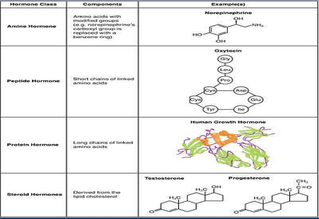
Steroid vs. Protein Hormones: How Signal Type Determines Physiologic Outcome
 We must distinguish steroid hormones from protein hormones because their signaling architectures dictate therapeutic approaches.
- Steroid hormones (e.g., estrogen, progesterone, testosterone, cortisol) are lipophilic and cross cell membranes. They bind intracellular nuclear receptors (e.g., ERα/ERβ for estrogen; AR for testosterone; PR for progesterone), then modulate gene transcription via hormone response elements. This influences mRNA transcription and protein synthesis, which can take hours to days to manifest. Steroid hormones also have rapid non-genomic effects through membrane-associated receptors, but their primary impact is transcriptional.
- Protein hormones (e.g., insulin, leptin, GLP-1) bind to membrane receptors and initiate second-messenger cascades—kinases, G-protein signaling, cAMP, Ca2+ flux—that alter cellular behavior rapidly. These effects are immediate and can be modulated by receptor density, post-receptor signaling integrity, and inflammatory states.
Clinical implication: When we optimize steroid hormone levels, we often engage gene expression programs that refine tissue architecture, enzyme production, mitochondrial function, and receptor abundance. When we modulate protein hormones (through nutrition, incretin therapies, or reducing inflammatory signaling), we alter the acute flux of energy and appetite signals. The two classes overlap in effects but differ fundamentally in their kinetics and levers.
Mitochondrial Health and Metabolic Flexibility: Why Energy Systems Break Down
Patients often ask, “Why do I feel tired despite eating clean and exercising?” The answer usually lies in mitochondrial function and metabolic flexibility.
- In insulin resistance, skeletal muscle’s ability to use glucose is diminished. This pushes fuel toward hepatic lipogenesis and adipose storage. The mitochondrial enzymes involved in beta-oxidation and oxidative phosphorylation are downregulated; ROS increases; mitochondrial DNA may become damaged; and biogenesis slows.
- AMPK and PGC-1α are central regulators. Lifestyle approaches that stimulate AMPK (through exercise intensity variation, caloric timing, polyphenols, and occasionally cold exposure) and PGC-1α (through endurance training and specific nutrient cues) can restore mitochondrial biogenesis and improve glycolytic-oxidative balance.
- Clinical reasoning for treatments: We deploy exercise prescriptions (resistance training for muscle mass and insulin sensitivity; zone 2 aerobic work for mitochondrial efficiency; high-intensity intervals sparingly to stimulate VO2max without overshooting stress) because they directly remodel mitochondrial networks and improve glucose disposal.
Example: A patient with Hemoglobin A1c of 5.7–6.4% (prediabetes) and elevated fasting insulin benefits from structured resistance training 3 days/week, plus 2–3 days of low-intensity aerobic sessions. We anchor meals around protein sufficiency and fiber density to support satiety and muscle repair, while moderating ultra-processed carbohydrates that spike insulin. Over 12–16 weeks, one can measure reduced HOMA-IR, a decline in fasting insulin, and improved continuous glucose monitoring (CGM) time-in-range—objective evidence of improved metabolic flexibility.
Inflammation, Adipose Tissue, and Cytokine Signaling: The Immunometabolic Loop
Adiposity is not just an energy depot; it is an endocrine organ. Hypertrophic adipocytes become hypoxic, leading to HIF-1α release and amplifying inflammatory gene expression. Macrophages shift toward an M1 phenotype, secreting pro-inflammatory cytokines. This state drives:
- Leptin resistance: Elevated leptin fails to suppress appetite because hypothalamic signaling is impaired by inflammatory and endoplasmic reticulum stress.
- Adiponectin decline: We lose one of the few adipokines that enhance insulin sensitivity and fatty acid oxidation.
- Increased TNF-α and IL-6 impair insulin receptor signaling via serine phosphorylation of IRS proteins.
Clinical reason to act: Reducing adipose tissue inflammation—through weight loss achieved by muscle-centric training and improved diet composition; sleep optimization; stress modulation; and possibly omega-3 and polyphenol intake—restores receptor signaling. The aim is to break the loop: better insulin signaling reduces inflammation; reduced inflammation restores neuroendocrine sensitivity; and improved sensitivity enables healthier weight dynamics.
Circadian Biology, Cortisol Rhythms, and Nocturnal Tissue Regeneration
Patients often report fatigue, poor sleep, and weight plateaus. Circadian physiology provides the framework:
- In healthy physiology, cortisol peaks in the early morning (the cortisol awakening response) and falls across the day, reaching its lowest point at night. Nocturnal low cortisol permits tissue regeneration, bone marrow dynamics, hepatic repair, and neuroplasticity.
- Testosterone and growth hormone pulses are aligned with sleep architecture. Disrupting sleep through late-night blue light exposure, irregular bedtimes, or stimulating hormones at night interferes with regenerative phases. I emphasize: nighttime is for repair; daytime is for performance.
Clinical application: I advise patients to remove televisions from the bedroom, limit bright screens 1–2 hours before bed, and align feeding with daylight. For patients on testosterone therapy, I avoid dosing patterns that spike levels at night. The goal is to preserve nocturnal anabolic-catabolic balance so skin, liver, bone marrow, and neural tissues can renew.
Estrogen Physiology: Skeletal Muscle, Brown Fat, Appetite, and Insulin Sensitivity
We must dispel myths: estrogen is not simply a “female reproductive hormone.” It is a metabolic regulator with systemic benefits in men and women.
- Estrogen (especially estradiol) enhances GLUT4-mediated glucose uptake in skeletal muscle, improves insulin sensitivity, and supports lipid oxidation.
- Estrogen influences brown adipose tissue (BAT) activation and thermogenesis, increasing energy expenditure. It modulates free fatty acid flux and helps maintain healthy adipose tissue function.
- In the hypothalamus, estrogen supports anorexigenic signals, influencing neuropeptide Y (NPY) and leptin sensitivity, thereby restraining appetite. Estrogen deficiency—common in menopause—predisposes to leptin resistance, fat gain, and decreased energy expenditure.
- Estrogen influences the balance between estrogen receptor alpha/beta and regulates gene expression in the breast, brain, liver, and vascular tissues. It also modulates oxidative stress and mitochondrial function.
Clinical note: Men with obesity may have altered aromatase activity, shifting androgen-estrogen balance, while women in perimenopause/menopause experience declining estradiol with consequent metabolic changes. Therapy is individualized: dosing must consider SHBG, free hormone fractions, hepatic metabolism, gut conjugation, and specific risk profiles. The aim is to restore metabolic tone without overshooting, using bioidentical forms when appropriate and aligning with current evidence and guidelines.
Progesterone, Neuroprotection, and Appetite Regulation
Progesterone often gets simplified to a “supportive” hormone for estrogen. In reality, progesterone exerts neuroprotective, anxiolytic, and metabolic effects.
- It modulates GABAergic tone, supporting sleep and stress resilience. It can influence appetite and thermogenesis and may protect the vascular endothelium.
- In menopausal care, progesterone paired with estradiol can improve sleep architecture, stabilize mood, and modulate appetite signals—thus indirectly supporting weight management.
Clinical reasoning: In women with sleep fragmentation, cyclic anxiety, or perimenopausal insomnia, appropriately dosed micronized progesterone at night can support restorative sleep, aiding nocturnal tissue repair and lowering late-night cortisol. This choice is made on a case-by-case basis, balancing benefits with breast and cardiovascular risk assessments.
Testosterone, Muscle, Metabolism, and Managing Risks
Testosterone is central to muscle mass, mitochondrial function, insulin sensitivity, and lipid metabolism in both sexes.
- In men, low testosterone correlates with increased adiposity, insulin resistance, dyslipidemia, and reduced aerobic capacity. In women, carefully managed testosterone can enhance lean mass, energy, and libido.
- Fear of cancer has historically overshadowed nuanced discussion of testosterone’s benefits and risks. Modern data suggest individualized, measured approaches minimize risks while supporting metabolic health.
Key clinical concern: Secondary polycythemia in testosterone therapy.
- Mechanism: Androgens can stimulate erythropoiesis, partly through renal EPO signaling and marrow stimulation. Elevated hematocrit increases viscosity, raising thrombotic risk.
- Protocol: Obtain baseline and periodic CBC (hematocrit/hemoglobin). If symptomatic (shortness of breath, headaches, dizziness), consider dose reduction, interval change, formulation shift (e.g., from injectable peaks to more stable delivery), or therapeutic phlebotomy when indicated. Evaluate sleep apnea, dehydration, and comorbidities that exacerbate erythrocytosis.
- Dosing rationale: Avoid large once-weekly spikes that disrupt nocturnal physiology. Favor dosing strategies that preserve daytime anabolic signaling while allowing nighttime lows for tissue regeneration. This aligns testosterone kinetics with circadian biology.
SHBG, Free Hormone Fractions, and Receptor-Level Nuance
A crucial concept: Symptoms do not always match total hormone levels because Sex Hormone-Binding Globulin (SHBG) and receptor sensitivity matter.
- SHBG binds estradiol and testosterone, shaping free and bioavailable hormone levels. In obesity and insulin resistance, SHBG often declines, altering free hormone availability and feedback loops.
- Obesity and inflammation can alter receptor expression, the balance of co-activators and co-repressors, and membrane trafficking, creating functional hypogonadism or functional estrogen deficiency even when total values appear “normal.”
- Clinical principle: Individualize. When a woman’s total testosterone is 110 ng/dL, do not judge by reference ranges alone. Consider SHBG, DHEA-S, estradiol, free testosterone, LH/FSH, thyroid status, liver function, and clinical symptoms. Reference ranges represent populations often burdened by obesity and diabetes; optimal physiological windows differ.
Gut Microbiome, Estrobolome, and Hormone Conjugation
We cannot ignore the gut microbiome.
- The estrobolome—bacterial genes capable of deconjugating estrogens via beta-glucuronidase—affects circulating estrogen levels. Dysbiosis can lead to aberrant estrogen recirculation or elimination, impacting metabolic tone, breast tissue signaling, and mood.
- A healthy gut supports Phase II conjugation and enterohepatic cycling; dysbiosis undermines these functions and inflames the intestinal barrier, allowing LPS endotoxemia to worsen insulin resistance and hepatic stress.
Clinical reasoning:
- Support gut integrity with fiber, polyphenols, fermented foods (when tolerated), and targeted probiotics where evidence supports specific strains. Avoid chronic, unnecessary antibiotics and reduce ultra-processed foods, which degrade microbial diversity.
- Monitor GI symptoms, stool patterns, and consider fecal testing when clinically indicated. Estrogen balance often improves when the gut is healthy, and inflammation is reduced.
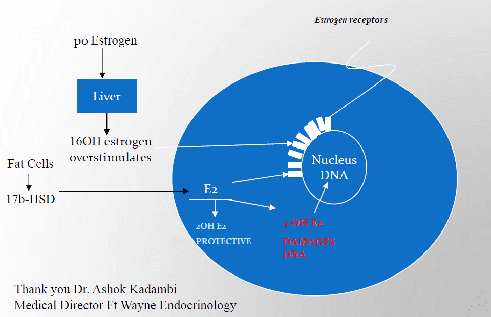
Phase I/II Detoxification, Hepatic Metabolism, and Hormone Clearance
Hormone balance depends on hepatic function:
- Phase I (cytochrome P450 enzymes) and Phase II (glucuronidation, sulfation, methylation) pathways metabolize steroid hormones and xenobiotics. If Phase I is upregulated and Phase II is sluggish, reactive intermediates may accumulate, exacerbating oxidative stress.
- Estrogen metabolism involves the 2-, 4-, and 16-hydroxylation pathways; the ratios of these metabolites influence tissue signaling and risk profiles. Clinical nuance includes nutritional co-factors supporting Phase II conjugation.
Clinical approach: We use nutrition and lifestyle strategies—adequate protein, B vitamins, magnesium, N-acetylcysteine, and sulforaphane-rich crucifers—to support conjugation. We monitor liver enzymes, consider alcohol intake, and manage comorbid NAFLD through weight loss and insulin sensitivity improvement.
Leptin, Appetite Regulation, and Hypothalamic Signaling
Leptin, produced by adipocytes, signals satiety to the hypothalamus. However, in obesity, leptin resistance develops:
- Inflammation, ER stress, and SOCS3 upregulation impair leptin receptor signaling, so elevated leptin fails to suppress appetite.
- Estrogen supports leptin sensitivity; declining estrogen worsens leptin resistance, increasing food intake and reducing energy expenditure.
Clinical intervention: Reduce inflammatory load, restore sleep and circadian alignment, and promote weight loss through muscle-centric strategies. Some patients benefit from time-restricted eating aligned with circadian rhythms—not as a fad, but as a way to improve glycemic excursions and normalize appetite signaling.
Why “Eat Less, Move More” Fails Without Metabolic Repair
Decades of public health advice focused on calorie restriction and generic exercise. Without repairing mitochondrial function and insulin/leptin signaling, many patients lose weight but also lose lean mass, worsen metabolic rate, and rebound.
- Clinical evidence shows that weight loss achieved through protein sufficiency, resistance training, and minimizing refined carbohydrate spikes preserves lean mass and enhances resting metabolic rate.
- Tracking metrics beyond weight—A1c, fasting insulin, CGM patterns, HOMA-IR, SHBG, ALT/AST (liver), CRP (inflammation)—gives a fuller picture. We tailor protocols to move these biomarkers toward healthy ranges.
Menopause, Andropause, and Individualized Hormone Replacement
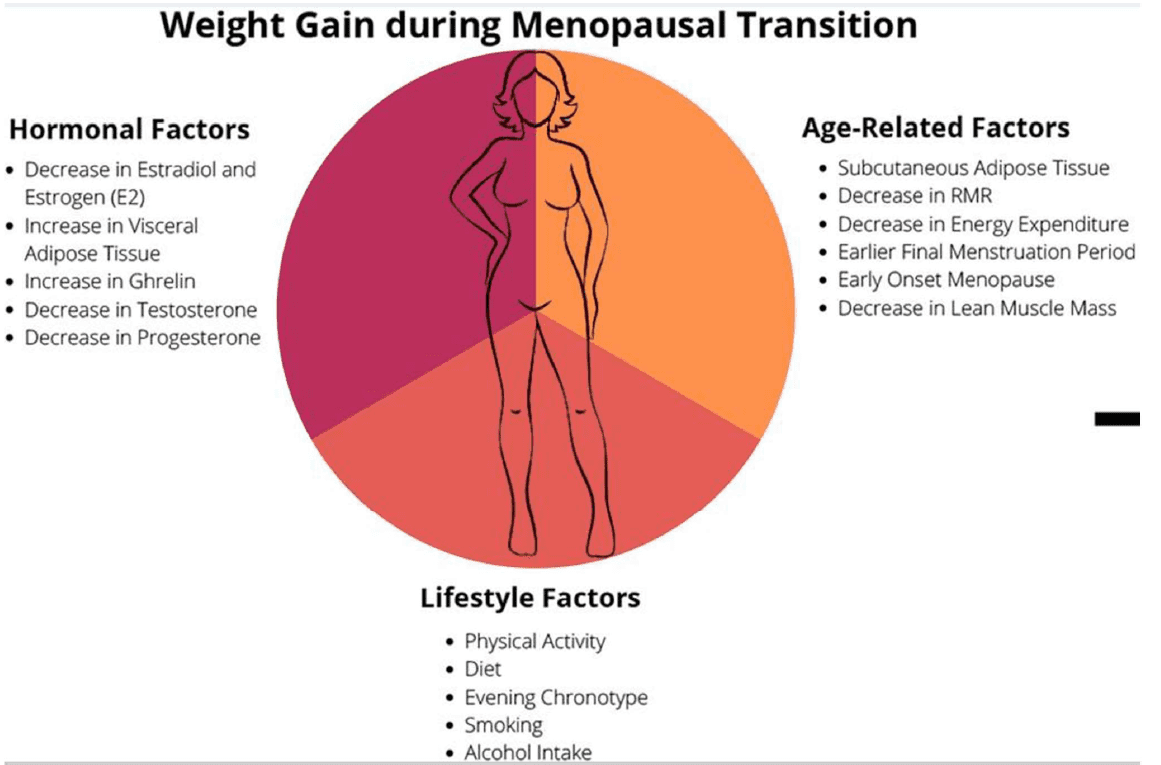 Hormone transitions are physiologic, but the modern environment—stress, circadian disruption, ultra-processed diets, sedentary lifestyles—magnifies symptoms and metabolic risk.
- Women: Decline in estradiol alters thermogenesis, insulin sensitivity, brain energy metabolism, and bone turnover. Micronized progesterone can support sleep and neuroprotection; transdermal estradiol may reduce thrombotic risk versus oral routes, depending on the patient.
- Men: Andropause involves a gradual decline in testosterone, increased visceral fat, and reduced muscle mass. Address sleep apnea, insulin resistance, and inflammation before or alongside therapy. Monitor hematocrit, lipids, PSA, and clinical symptoms carefully.
Clinical rationale: Using bioidentical hormones can align better with receptor affinity and metabolism. Pellets may offer convenience but reduce dosing flexibility; injectables can spike; transdermals provide steadier levels. The choice hinges on the patient’s physiology, preferences, risks, and our ability to monitor and adjust.
Polycythemia in Testosterone Therapy: Monitoring and Management
I am often asked about the frequency and management of testosterone-induced secondary polycythemia.
- Frequency varies by dose, route, baseline marrow status, and comorbidities (e.g., sleep apnea).
- Management: Baseline CBC, re-evaluate at 3 months, 6 months, then annually (sooner if symptomatic). If hematocrit rises—especially >54%—consider dose reduction, route change, increased hydration, sleep apnea evaluation, and therapeutic phlebotomy when indicated. Maintain nighttime lows to support marrow homeostasis and avoid nocturnal surges that complicate regeneration cycles.
Clinical reasoning: The goal is to optimize metabolic and functional benefits without increasing risk. Testosterone should not be a blunt instrument; it must be integrated into a comprehensive plan that includes sleep, nutrition, training, and inflammation control.
Practical Biomarkers and Thresholds: A1c, Insulin, and Glycemic Variability
Patients often ask, “What A1c defines diabetes?” The common thresholds:
- A1c 5.7–6.4%: Prediabetes
- A1c ≥6.5%: Diabetes
These are population thresholds; they do not capture glycemic variability, postprandial spikes, or insulin dynamics.
Clinical implication: A patient with normal A1c but elevated fasting insulin and poor CGM time-in-range may already exhibit insulin resistance. Treat the physiology, not just the category. We intervene earlier—by improving diet composition, exercise specificity, sleep, and stress management—to prevent disease progression.
Exercise Physiology: Resistance, Aerobic, and Recovery in Metabolic Repair
Exercise is a metabolic therapy when applied strategically:
- Resistance training increases muscle mass and GLUT4 expression, boosting insulin sensitivity and resting metabolic rate.
- Zone 2 aerobic training enhances mitochondrial density, fat oxidation, and lactate clearance—repairing metabolic flexibility.
- High-Intensity Interval Training (HIIT) can raise VO2max and insulin sensitivity in trained individuals; use judiciously to avoid excessive stress in fragile metabolic states.
- Recovery is essential. Over-exercising without sleep and nutrition degrades performance hormones and elevates cortisol.
Clinical reasoning: I prescribe individualized routines—e.g., 3 days of resistance, 2 days of Zone 2, optional 1 day of light intervals—paired with protein targets (1.2–1.6 g/kg/day in many adults, adjusted for kidney function and clinical context), fiber-rich carbohydrates, and healthy fats. We measure outcomes with performance, body composition, and lab markers.
Nutrition Science: Protein, Fiber, Carbohydrate Quality, and Meal Timing
Nutrition must serve physiology:
- Protein sufficiency supports muscle anabolism, satiety, and thermogenesis. Older adults often under-eat protein, accelerating sarcopenia.
- Fiber improves glycemic control, feeds beneficial microbiota, and reduces energy density.
- Carbohydrate quality matters: minimize refined sugars and ultra-processed starches that spike insulin and accelerate lipogenesis. Favor whole-food carbohydrates with intact fibers.
- Meal timing aligned with circadian rhythms—front-loading calories earlier in the day and avoiding late-night eating—supports cortisol rhythms, sleep, and glycemic control.
Clinical reasoning: These adjustments reduce postprandial hyperglycemia, enhance daytime energy, and support nocturnal regeneration. Patients report improved sleep, stable moods, and sustainable weight loss.
The Family Lens: Generational Risk and Early Intervention
I have watched families struggle as diabetes complications accumulate. In one example, both parents suffered severe diabetic complications. The adult child presented with multi-year labs showing progression toward insulin resistance. The standard advice they received—“exercise more and come back”—missed the urgency.
Clinical approach:
- We treat elevated risk with early, aggressive lifestyle and biomarker monitoring: A1c, fasting insulin, CGM, lipid subfractions, liver enzymes, SHBG, and CRP.
- We educate about mitochondrial and hormonal physiology to motivate sustained behavior change.
- We deploy structured training, nutrition, and sleep protocols, then personalize as biomarkers shift.
Outcome: Over months, we see improved insulin sensitivity, reduced visceral adiposity, and improved energy. This is prevention—anchored in physiology.
Managing Estrogen Fear and Weight Concerns in Women
Many women fear hormone therapy due to cancer risk or weight gain. We must clarify:
- Transdermal estradiol has different risk characteristics than oral therapy, particularly in thrombotic risk.
- Proper dosing, pairing with progesterone for endometrial protection (in women with a uterus), and careful screening reduce risks.
- Weight gain during menopause is often driven by declining estrogen’s effects on brown fat, appetite signaling, and insulin sensitivity—not by estrogen therapy itself. Therapy, aligned with lifestyle, can help restore metabolic tone and improve body composition.
Clinical reasoning: We individualize protocols, communicate risks and benefits transparently, and prioritize patient concerns. When a woman worries about weight, we integrate resistance training and protein sufficiency to protect lean mass while using hormone support judiciously.
Androgen Therapy in Women: Individualized, Measured, and Monitored
Women sometimes benefit from androgens for libido, energy, and lean mass. We attend closely to:
- Dose and formulation, starting low, monitoring symptoms and labs (total and free testosterone, SHBG, DHEA-S).
- Avoiding supraphysiologic levels that provoke acne, hair loss, or virilization. Clarify that reference ranges derive from populations with variable health; the target is functional well-being within a safe physiologic window.
Clinical reasoning: When properly individualized, androgen therapy in women can improve quality of life. We reassess frequently and adjust.
Sleep, Blue Light, and Neuroplasticity: Protecting the Regenerative Window
I routinely advise patients to remove TVs from the bedroom. It’s not an aesthetic preference; it’s physiology.
- Evening blue light suppresses melatonin, blunts growth hormone pulsatility, and elevates cortisol, impairing neuroplasticity and tissue regeneration.
- Patients who “feel relaxed” watching TV at night often suffer poor sleep architecture and morning fatigue. Over time, this worsens insulin resistance and weight gain.
Clinical reasoning: We choose behaviors that support regenerative biology—dark, cool bedrooms; consistent sleep schedules; evening wind-down routines; minimal alcohol; and aligned dosing of medications/hormones to avoid nocturnal stimulation.
Individual Variation and Receptor Dynamics: Why Symptoms Outpace Numbers
It bears repeating: hormone therapy is never “by the numbers” alone. Receptor sensitivity, co-factor availability, membrane fluidity, and post-receptor signaling all modulate outcomes. Someone can have “normal” hormone levels and significant symptoms if receptors are downregulated or signaling pathways are inflamed.
Clinical approach:
- Treat the patient, not just the paper. Integrate objective biomarker changes with subjective symptom improvements and functional outcomes (energy, sleep, mood, performance).
- Use trial periods with structured follow-up, adjusting based on response. Keep safety labs
Practical Questions Clinicians Ask: Superphysiologic Levels and Reference Ranges
“Is a total testosterone of 110 ng/dL in a woman acceptable?” The answer depends on:
- SHBG levels, free testosterone, DHEA-S, estradiol balance, clinical symptoms, and risk factors.
- “Superphysiologic” is not a fixed number; it is a physiological assessment relative to receptor expression and systemic effects.
- Reference ranges represent mixed populations, often metabolically unhealthy. Clinicians must target functional optimization within safety.
Clinical reasoning: I often use pellets initially for certain patients due to convenience and adherence, while acknowledging reduced flexibility. Over time, we may shift to modalities with finer control if needed.
Case Management: Secondary Polycythemia and Testosterone
When I encounter symptomatic erythrocytosis—patients complain of shortness of breath, headaches, or dizziness—I:
- Recheck CBC immediately, compare to baseline, and assess sleep, hydration, altitude exposure, and underlying pulmonary or renal conditions.
- Reduce dose, adjust route, and time dosing to minimize nocturnal peaks. Consider phlebotomy when indicated, especially with hematocrit exceeding safe thresholds.
- Continue addressing menopause/andropause physiology and coexisting metabolic conditions; don’t abandon testosterone reflexively—optimize it.
Implementation Roadmap: From Assessment to Action
- Baseline Assessment
- History: sleep, stress, appetite, weight trajectory, exercise, family risk.
- Labs: A1c, fasting insulin, CGM (optional), lipid panel, CRP, ALT/AST, SHBG, total/free testosterone, estradiol, progesterone (as applicable), CBC, TSH/T3/T4 (selected cases), vitamin D.
- Body composition: DXA or bioimpedance.
- Sleep study if symptoms suggest apnea.
- Initial Interventions
-
- Exercise: resistance 3x/week; Zone 2 aerobic 2–3x/week; optional light intervals.
- Nutrition: prioritize protein and fiber-rich whole foods; reduce ultra-processed carbs; align meal timing with daylight.
- Sleep hygiene: consistent schedule, dark room, blue-light restrictions, stress-reduction practices.
- Hormone Modulation (When Indicated)
-
- Women: consider transdermal estradiol, micronized progesterone; assess androgen support cautiously.
- Men: evaluate testosterone therapy with rigorous monitoring; align dosing to circadian biology; screen for polycythemia risk.
- Gut and Liver Support
-
- Fiber, polyphenols, fermented foods (as tolerated).
- Support Phase II with nutrient co-factors, moderate alcohol; monitor liver enzymes.
- Monitoring and Adjustment
-
- Reassess symptoms and labs at 8–12 weeks; refine exercise, nutrition, and dosing.
- Long-term: maintain quarterly-to-semiannual check-ins and an annual comprehensive lab review.
Debunking “Supplements Don’t Work”: Evidence-Based Nutraceutical Strategy
A common article once claimed most supplements “don’t work.” The truth is nuanced:
- Blanket dismissal ignores specific evidence. Omega-3s may improve triglycerides and inflammatory markers; vitamin D can affect bone and immune function; magnesium influences insulin sensitivity and sleep; and creatine supports muscle performance. Not all supplements fit all patients, and quality/dosing matters.
- Clinical reasoning: Use targeted nutraceuticals—e.g., magnesium glycinate for sleep and insulin sensitivity, creatine for muscle, omega-3 for triglycerides/inflammation—validated by patient-specific goals and labs. Avoid “shotgun” supplementation.
Real-World Barriers: Social Media, Celebrity Advice, and Mixed Messages
Patients often get their “medical advice” from celebrities or influencers. While intent may be good, advice is usually incomplete, non-individualized, and unmoored from physiology and lab data.
Clinical strategy:
- Empower patients with education. Explain why physiology matters—mitochondria, hormones, circadian rhythms—so they can discern quality guidance.
- Provide handouts and links customized to your practice. Encourage patients to track meaningful metrics and use tools such as CGM devices under clinical guidance.
The Vicious Cycle: Obesity, Hypogonadism, and Receptor Resistance
In obesity:
- Aromatase activity in adipose tissue alters androgen-estrogen balance.
- Inflammation reduces gonadal steroidogenesis and receptor sensitivity, causing hypogonadism in men and altered androgen balance in women.
- This worsens adiposity by reducing muscle mass and metabolic rate, fueling the cycle.
Clinical reasoning: Break the cycle by prioritizing muscle anabolism, reducing inflammation, restoring sleep, and considering hormone support when indicated. Receptors often recover as weight and inflammation decline.
Why Nighttime Testosterone Spikes Disrupt Regeneration
Nocturnal physiology is delicately tuned:
- High nighttime testosterone can disturb sleep architecture and reduce growth hormone pulses, interfering with skin, liver, bone marrow, and retinal
- Dosing that produces large peaks should be scheduled to avoid nighttime surges. This supportive approach respects circadian biology and preserves nocturnal tissue repair.
Safety Culture: Start Low, Go Slow, Monitor Always
I was trained to “start low,” titrate, and monitor—especially in long-standing hormone therapy cases:
- Measure baseline, then adjust at regular intervals—3, 6, 12 months—based on symptoms, labs, and function.
- This reduces the risk of overshooting and catches issues like erythrocytosis
Clinical reflection: This is the safest route to durable, high-quality outcomes in hormone therapy.
Personalized Medicine: Beyond Populations and Averages
Reference ranges describe populations—many obese, diabetic, and inflamed. Personalized medicine honors the individual by:
- Integrating biomarkers with clinical context and patient goals.
- Considering genetic, epigenetic, and microbiome
- Building sustainable protocols that patients can execute—exercises they enjoy, foods they can afford and prepare, sleep routines they can maintain.
Summary
Insulin resistance is the foundational disturbance driving modern chronic disease. It disrupts mitochondrial efficiency, inflames adipose tissue, and causes neuroendocrine dysfunction. Steroid and protein hormones act through distinct pathways, and recognizing their differences guides therapy. Estrogen supports insulin sensitivity, BAT activity, and appetite control; progesterone aids sleep and neuroprotection; testosterone sustains muscle metabolism but must be managed to prevent polycythemia and circadian disruption.
Restoring health requires aligning interventions with physiology: resistance and Zone 2 training to rebuild mitochondria and muscle; protein sufficiency and fiber-rich diets to support satiety and microbiome function; sleep hygiene to preserve nocturnal regeneration; and individualized hormone therapy with meticulous monitoring. SHBG, free hormone fractions, receptor dynamics, and gut-liver conjugation are essential nuances in care. We move beyond simplistic advice and deploy evidence-based, patient-centered strategies that repair the underlying metabolic machinery.
Unlocking the Secrets of Inflammation: Integrative Medicine Approach- Video
Conclusion
Treating metabolic and hormonal disorders demands a systems approach. When we respect circadian biology, restore mitochondrial function, reduce inflammation, and, when appropriate, carefully modulate hormones, patients regain metabolic flexibility, energy, and resilience. The difference between failure and success often hinges on the depth of our understanding and the precision of our protocols. My commitment is to present modern, translational research in a clinically actionable format—so patients and clinicians can collaborate toward outcomes that endure.
Key Insights
- Insulin resistance underlies most non-communicable chronic diseases; repairing it is priority one.
- Hormones signal via fundamentally different architectures—steroids via gene transcription, proteins via membrane cascades—requiring tailored interventions.
- Estrogen enhances skeletal muscle glucose uptake, BAT function, and appetite regulation; its decline increases metabolic risk.
- Testosterone supports muscle and metabolism across sexes but needs careful dosing to prevent polycythemia and nighttime disruption.
- SHBG, free hormone fractions, receptor sensitivity, and microbiome-driven conjugation explain why “normal” labs can coexist with symptoms.
- Circadian alignment—daytime performance, nighttime regeneration—is crucial; blue light and late dosing undermine repair.
- Muscle-centric training, combined with protein sufficiency, preserves metabolic rate and prevents rebound weight gain.
- Personalized care beats population averages; monitor, adapt, and integrate physiology into every clinical decision.
Keywords: metabolic health, insulin resistance, obesity, mitochondrial function, endocrinology, hormone therapy, estrogen, progesterone, testosterone, leptin, adipokines, neuroendocrine signaling, circadian biology, sleep, cortisol rhythm, menopause, andropause, bioidentical hormones, polycythemia, SHBG, GLP-1, adipose tissue, brown fat, muscle metabolism, mitochondrial biogenesis, inflammation, cytokines, chemokines, gut microbiome, estrobolome, hepatic metabolism, Phase I/II detoxification, clinical protocols, lifestyle medicine, exercise physiology, nutrition science, translational research
References:
- DeFronzo RA, Ferrannini E. Insulin resistance: a multifaceted syndrome responsible for NIDDM, obesity, hypertension, dyslipidemia, and atherosclerotic cardiovascular disease. Diabetes Care.
- Hotamisligil GS. Inflammation and metabolic disorders. Nature.
- Petersen KF, Shulman GI. Pathogenesis of skeletal muscle insulin resistance. J Clin Invest.
- Mauvais-Jarvis F. Estrogen and metabolic homeostasis. Diabetes.
- Clegg DJ. Estrogen and central regulation of energy balance. Endocrinology.
- Handelsman DJ. Testosterone therapy guidelines and erythrocytosis management. J Clin Endocrinol Metab.
- Boden G. Effects of insulin and free fatty acids on glucose metabolism. Diabetes.
- Paoli A. Ketogenic diets and physiological implications; circadian meal timing literature.
- Buxton OM, Czeisler CA. Circadian misalignment and metabolic risk. Proc Natl Acad Sci.
- Tremblay A, Simoneau JA. Exercise, mitochondrial function, and metabolic flexibility. Sports Med.
- Sonnenburg ED, Sonnenburg JL. Microbiota, fiber, and metabolic health. Cell Metab.
- Santoro N. Menopause and metabolic syndrome. Menopause.
- Veldhuis JD. Pulsatile hormone secretion and sleep. Endocr Rev.
Disclaimer:
- This educational post is not medical advice. It is for informational purposes only.
- All individuals must obtain recommendations for their personal situations from their own qualified medical providers.
Post Disclaimer
General Disclaimer, Licenses and Board Certifications *
Professional Scope of Practice *
The information herein on "Hormone Physiology Tip for Weight Management" is not intended to replace a one-on-one relationship with a qualified health care professional or licensed physician and is not medical advice. We encourage you to make healthcare decisions based on your research and partnership with a qualified healthcare professional.
Blog Information & Scope Discussions
Welcome to El Paso's Premier Wellness and Injury Care Clinic & Wellness Blog, where Dr. Alex Jimenez, DC, FNP-C, a Multi-State board-certified Family Practice Nurse Practitioner (FNP-BC) and Chiropractor (DC), presents insights on how our multidisciplinary team is dedicated to holistic healing and personalized care. Our practice aligns with evidence-based treatment protocols inspired by integrative medicine principles, similar to those on this site and on our family practice-based chiromed.com site, focusing on naturally restoring health for patients of all ages.
Our areas of multidisciplinary practice include Wellness & Nutrition, Chronic Pain, Personal Injury, Auto Accident Care, Work Injuries, Back Injury, Low Back Pain, Neck Pain, Migraine Headaches, Sports Injuries, Severe Sciatica, Scoliosis, Complex Herniated Discs, Fibromyalgia, Chronic Pain, Complex Injuries, Stress Management, Functional Medicine Treatments, and in-scope care protocols.
Our information scope is multidisciplinary, focusing on musculoskeletal and physical medicine; wellness; contributing etiological viscerosomatic disturbances within clinical presentations; associated somato-visceral reflex clinical dynamics; subluxation complexes; sensitive health issues; and functional medicine articles, topics, and discussions.
We provide and present clinical collaboration with specialists from various disciplines. Each specialist is governed by their professional scope of practice and licensure jurisdiction. We use functional health & wellness protocols to treat and support care for musculoskeletal injuries or disorders.
Our videos, posts, topics, and insights address clinical matters and issues that directly or indirectly relate to our clinical scope of practice.
Our office has made a reasonable effort to provide supportive citations and has identified relevant research studies that support our posts. We provide copies of supporting research studies upon request to regulatory boards and the public.
We understand that we cover matters that require an additional explanation of how they may assist in a particular care plan or treatment protocol; therefore, to discuss the subject matter above further, please feel free to ask Dr. Alex Jimenez, DC, APRN, FNP-BC, or contact us at 915-850-0900.
We are here to help you and your family.
Blessings
Dr. Alex Jimenez DC, MSACP, APRN, FNP-BC*, CCST, IFMCP, CFMP, ATN
email: [email protected]
Multidisciplinary Licensing & Board Certifications:
Licensed as a Doctor of Chiropractic (DC) in Texas & New Mexico*
Texas DC License #: TX5807, Verified: TX5807
New Mexico DC License #: NM-DC2182, Verified: NM-DC2182
Multi-State Advanced Practice Registered Nurse (APRN*) in Texas & Multi-States
Multi-state Compact APRN License by Endorsement (42 States)
Texas APRN License #: 1191402, Verified: 1191402 *
Florida APRN License #: 11043890, Verified: APRN11043890 *
Colorado License #: C-APN.0105610-C-NP, Verified: C-APN.0105610-C-NP
New York License #: N25929, Verified N25929
License Verification Link: Nursys License Verifier
* Prescriptive Authority Authorized
ANCC FNP-BC: Board Certified Nurse Practitioner*
Compact Status: Multi-State License: Authorized to Practice in 40 States*
Graduate with Honors: ICHS: MSN-FNP (Family Nurse Practitioner Program)
Degree Granted. Master's in Family Practice MSN Diploma (Cum Laude)
Dr. Alex Jimenez, DC, APRN, FNP-BC*, CFMP, IFMCP, ATN, CCST
(Board Certified: Family Practice Nurse Practitioner—Multistate)*
(Licensed Nurse Practitioner & Chiropractor - Multistate)*
Clinical Director
Digital Business Card
Dr. Maria Cardenas, MD
(Board Certified: Internal Medicine)
(Licensed Medical Doctor)
Medical Director, Clinical Director & Collaborative Physician
NPI # 1164426749
MD License #: J2933
Licenses and Board Certifications:
MD: Medical Doctor
DC: Doctor of Chiropractic
APRNP: Advanced Practice Registered Nurse
FNP-BC: Family Practice Specialization (Multi-State Board Certified)
RN: Registered Nurse (Multi-State Compact License)
CFMP: Certified Functional Medicine Provider
MSN-FNP: Master of Science in Family Practice Medicine
MSACP: Master of Science in Advanced Clinical Practice
IFMCP: Institute of Functional Medicine
CCST: Certified Chiropractic Spinal Trauma
ATN: Advanced Translational Neutrogenomics
Memberships & Associations:
TCA: Texas Chiropractic Association: Member ID: 104311
AANP: American Association of Nurse Practitioners: Member ID: 2198960
ANA: American Nurse Association: Member ID: 06458222 (District TX01)
TNA: Texas Nurse Association: Member ID: 06458222
NPI: 1205907805
| Primary Taxonomy | Selected Taxonomy | State | License Number |
|---|---|---|---|
| No | 111N00000X - Chiropractor | NM | DC2182 |
| Yes | 111N00000X - Chiropractor | TX | DC5807 |
| Yes | 363LF0000X - Nurse Practitioner - Family | TX | 1191402 |
| Yes | 363LF0000X - Nurse Practitioner - Family | FL | 11043890 |
| Yes | 363LF0000X - Nurse Practitioner - Family | CO | C-APN.0105610-C-NP |
| Yes | 363LF0000X - Nurse Practitioner - Family | NY | N25929 |
Dr. Alex Jimenez, DC, APRN, FNP-BC*, CFMP, IFMCP, ATN, CCST
(Board Certified: Family Practice Nurse Practitioner—Multistate)*
(Licensed Nurse Practitioner & Chiropractor - Multistate)*
Clinical Director
Digital Business Card
Dr. Maria Cardenas, MD
(Board Certified: Internal Medicine)*
(Licensed Medical Doctor)*
Medical Director, Clinical Director & Collaborative Physician
NPI # 1164426749
MD License #: J2933
📆 Schedule Appointment: Schedule 24/7 (Click Here)