


Table of Contents
By Dr. Jimenez, DC, FNP-APRN
Introduction: A New Paradigm for Weight Management
Welcome to this educational post, where we will delve into the intricate and multifaceted world of weight management, moving far beyond the outdated, often ineffective mantra of “eat less, exercise more.” As a clinician with 4 decades of experience and dual credentials as a Doctor of Chiropractic (DC) and a Family Nurse Practitioner (FNP-APRN), my practice is built on integrating diverse medical perspectives to create a holistic, evidence-based understanding of human physiology. The reality is that sustainable weight loss is not about willpower alone; it is a complex interplay of genetics, physiology, environment, and lifestyle. In this comprehensive discussion, I will share insights gleaned from my practice and the latest findings from leading researchers, presenting a modern, evidence-based framework for understanding and addressing weight gain. We will explore why traditional weight-loss methods often fail and why the advent of new pharmaceuticals, while promising, requires a nuanced, holistic approach to ensure long-term success and health.
This post will begin by deconstructing the common pitfalls of conventional weight loss, particularly the detrimental loss of lean muscle mass that often accompanies rapid weight reduction. We will then introduce the Pratt-Wooten approach, a philosophy centered on patient responsibility and a deep understanding of the foundational pillars of health: nutrition, exercise, and sleep. From there, we will embark on a deep dive into the physiological systems that govern our weight. We will explore the profound impact of epigenetics, understanding how our daily choices and environmental exposures can alter gene expression, influencing everything from our stress response to our predisposition to weight gain. A central theme will be the interconnectedness of our body’s systems—a concept known as systems biology. We will examine how the cortisol bone connects to the insulin bone, which in turn connects to the thyroid bone, illustrating how stress, blood sugar dysregulation, and hormonal imbalances are inextricably linked.
We will thoroughly investigate key physiological disruptors, including chronic stress and its effect on the cortisol curve, the critical role of glucose-insulin regulation, and the emerging science of the gut microbiome and its influence on inflammation and metabolism. We will also address the unique challenges faced by different populations, such as post-menopausal women who experience a natural shift towards a pro-inflammatory state. Furthermore, we will discuss the often-overlooked impact of environmental toxins on metabolic function and the critical importance of micronutrients, with a special focus on magnesium, a mineral deficiency strongly linked to metabolic syndrome. Finally, we will contextualize this knowledge within the current landscape of weight loss therapies, including the widespread use of GLP-1 receptor agonists. We will discuss how to use these powerful tools responsibly, emphasizing the need for a comprehensive lifestyle program to support patients, mitigate side effects such as muscle and nutrient loss, and lay a foundation for lasting health. This post is designed to provide you with a robust, scientifically-grounded understanding of weight management, empowering you to think critically and holistically about this crucial aspect of health and well-being.
Deconstructing the Weight Loss Paradigm: Beyond the Scale
For the past 40 years, I’ve dedicated my career to helping patients achieve and maintain a healthy weight. A recurring theme I’ve observed is the frustration and ultimate failure that come from a simplistic approach to a complex problem. When individuals embark on a weight-loss journey, especially through popular medical weight-loss programs, they often see initial success on the scale. It’s not uncommon for them to lose three to four pounds quickly. However, a critical question we must ask is: what kind of weight are they losing?
In many cases, a significant portion of this initial loss is not fat, but precious lean muscle mass. This is a critical problem. Lean muscle is our metabolic engine. It burns calories even at rest, supports our skeletal structure, and is vital for strength and mobility. Losing it is counterproductive to long-term health and weight management. The issue is that muscle is dense and metabolically active; losing it makes it progressively harder to continue losing fat and significantly easier to regain weight once the intensive phase of a diet ends. This is the fundamental flaw in the “calories in, calories out” model when applied in isolation. It doesn’t account for the quality of the weight being lost.
My approach, which I’ve refined over decades and sometimes refer to as the Pratt-Wooten approach, is fundamentally about shifting the locus of control back to the individual. The ultimate goal is to move patients away from a reliance on a “pill for every ill” and toward taking profound responsibility for their own health. If a person doesn’t internalize this responsibility, their chances of long-term success are slim. The reality is that, in general, modern life encourages habits that lead to weight gain: we eat too frequently, our portion sizes are too large, we lead sedentary lives, and we consistently fail to get adequate, restorative sleep. These are the foundational pillars we must address.
To guide patients effectively, I utilize a comprehensive decision-tree rubric. This isn’t a one-size-fits-all plan but a systematic way of thinking that allows us to consider all the contributing factors. We review the patient’s history, current lifestyle, and metabolic markers, then decide on appropriate interventions, including lifestyle changes, nutritional support, or the judicious use of pharmaceutical agents. This structured approach ensures we are not just chasing symptoms but addressing the root causes of weight gain.
The Epigenetic Blueprint: How Lifestyle Shapes Your Genetic Destiny
One of the most powerful concepts in modern medicine is epigenetics. While we are born with a genetic code, our lifestyle and environment act as switches that can turn genes on or off. This means your daily choices directly impact your health trajectory.
Think about the developmental origins of health and disease. The environment you were exposed to in the womb, the stress levels your mother experienced, and her nutritional status all left an epigenetic imprint on you. This programming can influence your nerve health, your metabolic pathways, and even your behavioral responses to stress for the rest of your life. For example, some individuals have genetic variants that affect their dopamine pathways. Under stress, their brains don’t produce or process this “reward” neurotransmitter efficiently, leading them to seek external sources of pleasure and comfort, very often in the form of highly palatable, high-calorie foods. This isn’t a moral failing; it’s a biological predisposition. Understanding this requires taking a deep in-depth history. Has there been trauma or abuse? Is there a physiological reason why the body is craving certain nutrients or substances? By understanding these genetic predispositions, we can offer targeted support, perhaps through specific nutrients or therapies that help modulate dopamine levels, thereby helping someone overcome these powerful, biologically driven urges.
The Impact of Modern Life’s Disruptors
Understanding these epigenetic influences means we must also identify and address the modern-day disruptors that sabotage our health. I see this constantly in my practice. Consider the highly motivated individual who is desperate to lose weight. They might get up at 4:00 a.m. to attend a 5:00 a.m. boot camp, fueled by the belief that more exercise is always better. However, they don’t get to bed until 11:00 p.m. or later because of work, family, and household responsibilities. They are chronically sleep-deprived, running on stress hormones.
This scenario is a perfect storm for weight gain, not loss. The lack of sleep and intense early-morning exercise sends their cortisol—the primary stress hormone—skyrocketing. Chronically elevated cortisol disrupts blood sugar, promotes insulin resistance, and signals the body to store fat, particularly in the abdominal area. They are working against their own biology. One of the first and most crucial things we must do is get their stress and sleep under control. This is not easy, and it’s a highly individualized process, but it is non-negotiable for sustainable success.
The landscape of weight has changed dramatically. When I was growing up, seeing an overweight child was an anomaly. That child was often singled out and teased relentlessly. My own brother was that kid, and his weight eventually climbed to 466 pounds. I lived through that experience with him, and it profoundly shaped my professional path. Today, obesity is no longer an oddity; it’s an epidemic. This shift is a direct result of our changing environment, food supply, and lifestyle—all factors that exert powerful epigenetic pressures on our population.
The Interconnected Web of Health: A Systems Biology Approach
To truly understand and manage weight, we must adopt a systems biology approach. This means recognizing that the body is not a collection of separate parts but a deeply interconnected network. You can’t treat one system in isolation without affecting all the others. A common phrase I use to illustrate this is: “The cortisol bone is connected to the insulin bone, and the insulin bone is connected to the thyroid bone.”
Let’s break this down:
- Cortisol and Insulin: When you are under chronic stress, your body produces high levels of cortisol. Cortisol’s job is to prepare you for a “fight or flight” situation by mobilizing energy. It does this by telling your liver to release stored glucose into the bloodstream. This surge in blood glucose then triggers a release of insulin from the pancreas to shuttle that sugar into your cells. If this happens occasionally, it’s a normal adaptive response. But if you are chronically stressed, your cells are constantly being bombarded with insulin. Over time, they become resistant to its signal, a condition known as insulin resistance. Your pancreas has to work harder, pumping out even more insulin to get the job done. High insulin levels are a primary driver of fat storage. Therefore, elevated cortisol levels lead to decreased insulin sensitivity.
- Cortisol and Thyroid: High cortisol also interferes with thyroid function. Your thyroid gland produces a storage hormone called thyroxine (T4). For your metabolism to function optimally, T4 must be converted into the active thyroid hormone, triiodothyronine (T3). Chronic stress and high cortisol levels inhibit this crucial T4-to-T3 conversion. The result is a functional hypothyroidism, where your metabolic rate slows down, leading to fatigue, brain fog, and weight gain, even if your standard thyroid tests (like TSH) look normal.
- Cortisol and Gut Health: Stress also profoundly impacts your gut. High cortisol levels can increase intestinal permeability, also known as “leaky gut.” This allows undigested food particles and bacterial toxins to enter the bloodstream, triggering an immune response. This chronic, low-grade inflammation, originating in the enteric nervous system (the “brain in your gut”), can lead to the development of new food sensitivities and allergies, further fueling the inflammatory fire and contributing to metabolic dysfunction.
We are only as strong as our weakest link. In the world of “biohacking,” people often get confused and try to optimize everything at once. They might be taking dozens of supplements, doing cold plunges, and using infrared saunas without a clear, integrated strategy. This can backfire. A successful approach requires a complete, thoughtful plan that moves a person systematically through the healing process, teaching them to create a sustainable lifestyle that supports their unique biology.
The ultimate goal is not just weight loss but creating a life where maintaining a healthy weight feels natural and effortless. I have never met anyone who lost weight, kept it off, and was unhappy about it. The misery comes when they hit their goal weight, only to feel helpless as the pounds start creeping back on. This is what we must prevent.
The Multifactorial Nature of Obesity: Why “Eat Less, Move More” Fails
Obesity is a multifactorial condition. It’s not just about personal choices; it’s a complex interplay of various factors:
- Social and Environmental: I grew up in an environment where social life revolved around places like the local pizza inn. Our modern food environment is saturated with cheap, hyperpalatable, calorie-dense foods engineered to be addictive.
- Behavioral: Many of our eating habits are learned behaviors, acquired in childhood and reinforced over the years.
- Metabolic and Cellular: As we’ve discussed, hormonal imbalances, insulin resistance, and inflammation create a cellular environment that promotes fat storage.
- Lifestyle: Something as seemingly simple as working a night shift can have profound metabolic consequences. Eating at night goes against the natural circadian rhythm of our digestive and metabolic systems. For mammals, we are designed to eat during the day and fast at night. When a person’s job requires them to flip this cycle, we are working against a fundamental biological blueprint, and we must find creative strategies to help them mitigate the metabolic damage.
The statistics are staggering. It’s estimated that by the year 2030, 50% of the U.S. population will have obesity. We are already seeing the consequences. The National Institutes of Health (NIH) recognized years ago that a new strategic plan was needed for obesity, acknowledging that the simple advice of “eat less and exercise more” was failing to curb the epidemic. Yet, this is still the dominant message many people hear.
This advice might work for a 20-year-old with a resilient metabolism. But as we age, our hormonal milieu changes, our metabolic flexibility decreases, and our bodies become more resistant to weight loss. I’ve had countless patients who follow their diet and exercise plan meticulously for a week, only to step on the scale and find they’ve gained a pound. This isn’t a rare occurrence. It’s a deeply frustrating reality for many, and it happens because their program isn’t addressing the underlying whole-body metabolic dysfunction.
The Rising Tide of Diabetes and Metabolic Syndrome
The obesity epidemic is walking hand-in-hand with an epidemic of prediabetes and Type 2 diabetes. Currently, an astonishing 38% of the U.S. population is considered prediabetic. The COVID-19 pandemic appears to have accelerated this trend, with studies showing that a COVID-19 infection can increase the risk of developing diabetes by over 20%. I’ve seen this firsthand in my clinic: previously healthy, fit individuals who, after a bout with COVID (or in some cases, post-vaccination), experienced a sudden and dramatic shift in their metabolic health. This is likely due to profound changes in inflammatory cytokines and the microbiome.
The progression from prediabetes to diabetes is alarmingly common. Research indicates that up to 70% of individuals with prediabetes will eventually develop full-blown diabetes. We have robust data to quantify this risk. A large-scale Japanese study published in 2022, along with data from the Kaiser Permanente system, provides a clear picture. These studies converge on a frightening reality: your risk of developing diabetes within 10 years increases significantly with even small elevations in your fasting glucose.
- The Kaiser study suggests a 6% increased 10-year risk for every 1 mg/dL increase in fasting glucose above 84 mg/dL.
- The Japanese study found a 9% increased risk for every 1 mg/dL increase above 81 mg/dL.
Let’s put this into perspective. The standard lab range for “normal” fasting glucose often goes up to 99 mg/dL. If your fasting glucose is 99, you are just one point away from the official prediabetes diagnosis. According to this data, your 10-year risk of developing diabetes is already between 81% and 84%. This is based solely on glucose, without even considering other crucial markers like your triglyceride-to-HDL ratio, fasting insulin levels, or fructosamine (a marker of average blood sugar over the past 2-3 weeks).
We are not doing a good enough job of communicating the gravity of these numbers to our patients. A fasting glucose of 95 is not “good” or “normal”; it’s a warning sign of impending metabolic disaster. I saw this with my own father. At age 79, his fasting glucose was in the high 90s, and he had a 4- to 8-fold increased risk of gastrointestinal cancers associated with that level of dysglycemia. We intervened aggressively: we changed his diet, I introduced him to a treadmill, and we used targeted nutrients and injections. He lived to be 91. The point is that with the right interventions, we can change this trajectory. But people must first understand the severity of the situation. We have patients in their 50s with the kidney function (measured by eGFR) of an 80-year-old, and it’s directly linked to this chronic, low-level blood sugar dysregulation. It’s a silent and disturbing crisis.
The Weight Loss Industry: A Maze of Misinformation
The weight loss industry is a $66 billion behemoth, built largely on fleeting trends and misinformation. I’ve seen them all: the cabbage soup diet, the grapefruit diet, and countless others. These are not sustainable solutions. A concerning trend is the rise in unsupervised weight loss, with an estimated 80% of childhood weight-loss attempts occurring without medical supervision.
This has become particularly dangerous with the advent of research chemicals and the online black market for pharmaceuticals. You have individuals who are desperate, perhaps 50 or 70 pounds overweight, who go onto forums and Facebook pages to get advice on how to dose powerful medications. I’ve seen posts where someone asks, “Is this the right amount?” while holding up a syringe of a peptide, as if they were asking about a vitamin supplement. These are not supplements. These are potent drugs with specific half-lives and the potential to alter your physiology and gene expression profoundly. People are experimenting with daily micro-dosing protocols, fundamentally changing the pharmacokinetics of these drugs without any understanding of the long-term consequences. This is a recipe for disaster.
Meanwhile, anyone can hang a shingle and call themselves a weight loss expert. The sad reality is that only about 5% of people who lose significant weight manage to keep it off long-term. This is the statistic we must change.
Key Physiological Disturbances That Drive Fat Gain
To change that statistic, we must identify and address the core physiological disturbances that contribute to weight gain. Here is a summary of the key areas we must investigate in every patient:
- Hormonal Imbalances: This is the central hub. As we’ve discussed, imbalances in cortisol, insulin, and thyroid hormones are primary drivers. We also have to consider sex hormones. As a post-menopausal female’s estradiol levels decline, her body’s inflammatory setpoint shifts. Her immune cells, such as macrophages, shift from a predominantly anti-inflammatory state (M2) to a pro-inflammatory state (M1). This happens simply because the protective, anti-inflammatory signal of estradiol is gone. This is a crucial consideration in her weight loss journey.
- Chronic Nutritional Deficiencies: The single most important nutrient deficiency associated with metabolic syndrome (the trifecta of prediabetes, hypertension, and obesity) is magnesium. The NIH itself highlights this connection. It’s estimated that up to 77% of the population is functionally deficient in magnesium. Why? Our modern agricultural practices have depleted the soil of this vital mineral. Magnesium is the central atom in the chlorophyll molecule—it’s what makes green plants green. Even if people eat plenty of leafy green vegetables, the magnesium content is highly variable. In today’s world, it’s nearly impossible to get adequate magnesium without supplementation. It’s involved in over 300 enzymatic reactions in the body, many of which are critical for energy metabolism and insulin signaling. Other key nutrients include chromium and zinc, which are cofactors for the insulin receptor, and B vitamins like B1 (thiamine), which are essential for carbohydrate metabolism.
- Infections and Biotoxin Exposure: Chronic infections or exposure to biotoxins (such as mold) can induce a state of the Warburg effect (aerobic glycolysis). This is an inefficient metabolic state where cells produce energy without using oxygen, even when it’s available. Instead of generating 36+ ATP molecules from one glucose molecule through efficient mitochondrial respiration, the cell only generates 2 ATP. This is a survival state that cripples metabolic efficiency.
- Mitochondrial Dysfunction: Your mitochondria are the powerhouses of your cells. When damaged, they “leak” electrons, causing massive oxidative stress. The popular solution today is to “shoot up a bunch of NAD.” But if you have damaged mitochondria, simply jacking up NAD levels without addressing the underlying damage is like flooring the gas pedal in a car with a broken engine. You’re just driving more oxidative stress. The key is first to repair and reinvigorate the mitochondria.
- Gut Dysbiosis and Integrity: The health of your gut microbiome is paramount. An imbalance in gut bacteria (dysbiosis) and a compromised gut lining (“leaky gut”) are major drivers of chronic inflammation, which directly contributes to insulin resistance and fat storage. We all have a unique microbial “footprint,” and optimizing it is a key part of the weight loss journey.
- Impaired Detoxification: Our bodies are constantly exposed to environmental toxins. Arsenic, for example, is known to interfere with insulin regulation. Other heavy metals can disrupt thyroid function. If your body’s detoxification pathways are overwhelmed or inefficient, these toxins can accumulate, sabotaging your metabolic health.
- Medications: Many common medications can cause significant weight gain. SSRIs like sertraline are a classic example. A patient might be eating only 800 calories a day but still gain 40 pounds on the medication due to its metabolic effects. We must always consider the impact of a patient’s medication list.
- Physical Inactivity (The Modern Default): Our environment is engineered for inactivity. You can use a golf cart to get your mail from a 50-foot driveway. We have zero-turn-radius lawnmowers. These “innovations” rob us of the natural, daily movement our bodies are designed for. I closely track my own activity and exertion levels. I was shocked to find that mowing my lawn with a push mower generated a “strain score” on my fitness tracker that was as high as a structured weightlifting workout. We have to consciously re-integrate physical exertion into our lives. People are not getting their steps in or exerting themselves. This is a massive problem.
The Downward Spiral of Metabolic Dysfunction
When these systems are out of balance, a vicious cycle begins. It looks like this:
- Glucose control worsens, leading to more inflammation.
- Inflammation signals the body to store more visceral (belly) fat.
- Blood pressure rises, and blood lipids become dysregulated (dyslipidemia).
- The accumulation of belly fat further uncouples insulin receptor sensitivity, meaning your cells become even more resistant to insulin’s signal.
- You produce more lactate through inefficient metabolism, which makes you feel sore and fatigued.
- Your immune system becomes less effective.
- Cognitive function declines—that “brain fog” feeling.
- Sex hormone production plummets.
This is the downward spiral of metabolic syndrome, and it all starts with these interconnected disturbances.
A Note on GLP-1 Agonists and Nutritional Health
The new class of drugs, GLP-1 receptor agonists, has been a game-changer for many. However, they are not a magic bullet. It’s crucial to understand that these drugs don’t cause nutritional deficiencies directly. What happens is that they are so effective at suppressing appetite that people stop eating enough.
Obese individuals are often already nutrient-deficient. Then, you put them on a GLP-1 agonist, and their food intake plummets. Suddenly, they’re losing their hair. Why? They are not consuming enough protein. They are not getting the essential vitamins and minerals their body needs to function. You can’t build and maintain your body’s structures—hair, skin, nails, muscle, enzymes—without adequate protein. Many people on these medications lose their desire for foods like meat, chicken, or fish. So, a key part of our job is to figure out how to help them get that essential protein in, often through high-quality protein supplements and creative meal planning. We must be vigilant in monitoring and supplementing key nutrients, such as magnesium, chromium, zinc, and B vitamins, to support their metabolic machinery as they lose weight.
Losing weight is more than just calories in versus calories out. It’s about healthily changing body composition, addressing the multiple health implications of obesity, and creating a foundation of nutritional adequacy that will support a lifetime of health. This requires a comprehensive, thoughtful, and deeply personalized approach.
The Intricate Web of Metaflammation: Unraveling the Roots of Chronic Disease
In my clinical practice, I see the consequences of our modern lifestyle every single day. When we talk about weight gain, the conversation often defaults to a simple equation: calories in, calories out. While energy balance is a component, it’s a dramatically oversimplified view of a deeply complex biological process. The reality is that when we carry extra weight, we place significantly more pressure on our joints, a mechanical stress that can lead to pain and degeneration. But the more insidious problem lies at the cellular level. As weight and, consequently, Body Mass Index (BMI) increase, a cascade of metabolic disturbances is set in motion. For many of my patients, this is visibly reflected in rising blood pressure, a clear and immediate sign that the cardiovascular system is under strain.
The problem is deeply rooted in the quality and function of our adipose tissue, or fat. When your BMI exceeds 30, which marks the clinical definition of obesity, the physiology of your fat cells changes dramatically. We need to move beyond thinking of fat as a single entity and instead understand its different types. We have white adipose tissue (WAT), which primarily stores energy; brown adipose tissue (BAT), which is rich in mitochondria and actively burns energy to produce heat; and beige adipose tissue, a form of white fat that can be induced to behave more like brown fat. In a healthy metabolic state, the body can convert white fat into beige fat, which can then be activated to function like brown fat. This process is incredibly beneficial for burning excess calories and maintaining metabolic flexibility.
However, in a state of chronic over-nutrition and inflammation, this crucial conversion is blocked. You don’t get that healthy transition from beige to brown. Instead, your white adipose tissue proliferates and becomes dysfunctional. It becomes what I call “marbled” and “yellow”—it’s no longer a healthy, responsive tissue but an inflamed, inert storage depot.
Visualizing Insulin Resistance: The Concept of “Marbled” Muscle
I often use an analogy to help my patients visualize what’s happening inside their bodies, and while it may be a bit stark, it’s effective. Think about the difference between a lean cut of steak and a heavily marbled one. A DEXA scan (Dual-Energy X-ray Absorptiometry), which we use a tool to measure body composition, can provide similar insights into your tissues. It doesn’t just tell you how much muscle you have; it can give us an indication of the quality of that muscle—how lean it is.
When a person becomes insulin-resistant, their body’s ability to build and maintain lean muscle mass is severely compromised. Insulin is not just a blood sugar hormone; it’s a powerful anabolic hormone, meaning it’s essential for growth and repair, including muscle protein synthesis. When your cells stop responding properly to insulin, you effectively block the creation of new lean mass. Instead of building strong, functional muscle, the body begins to store fat within the muscle tissue itself, a condition known as intramyocellular lipids. This is the “marbling” I refer to. Your muscles become infiltrated with fat, making them less efficient at taking up glucose and further exacerbating the cycle of insulin resistance. You don’t want this unhealthy, inflamed, “white” tissue infiltrating your vital organs and muscles. The underlying inflammation and cellular senescence drive this process (the process by which cells age and stop dividing) that characterizes metabolic syndrome, making the entire system progressively more dysfunctional.
The Inflammatory Cascade: How Metaflammation Hijacks Insulin Signaling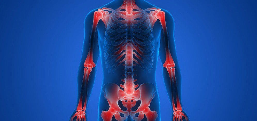
So, what is driving this process at the molecular level? It’s a phenomenon that researchers have termed metaflammation—a low-grade, chronic, systemic inflammation triggered by metabolic overload. When your fat cells become over-stuffed and dysfunctional, they begin to act like rogue immune cells, pumping out a host of inflammatory signaling molecules called cytokines.
Two of the most notorious culprits in this process are Interleukin-6 (IL-6) and Tumor Necrosis Factor-alpha (TNF-α). My own research and the work of leading immunologists have shown that when these cytokines are chronically elevated, they directly sabotage insulin signaling. They bind to receptors on the surface of your cells and trigger an intracellular cascade that essentially phosphorylates the insulin receptor on the wrong spot (serine phosphorylation instead of tyrosine phosphorylation). This conformational change makes the receptor unresponsive to insulin. It’s like putting the bad key in a lock; the signal can’t get through.
What is the body’s response to this blockade? The pancreas, sensing that glucose levels are not dropping, does the only thing it knows how to do: it pushes out more insulin. This state of high insulin levels is known as hyperinsulinemia. For a while, this brute-force approach works, but it comes at a cost. It further desensitizes receptors and places immense strain on the pancreas’s beta cells, which produce insulin.
This inflammatory state has far-reaching consequences. For example, we see conditions like alopecia (hair loss) linked to this systemic inflammation. Another hormone, leptin, which is supposed to signal satiety to the brain, becomes dysregulated. In a state of metaflammation, you develop leptin resistance: even with high leptin levels, your brain can’t hear the signal, so you keep feeling hungry.
Furthermore, this inflammatory environment leads to the breakdown of tissues, such as the myelin sheath that protects our nerves. The debris from this breakdown, which includes complex molecules like lipopolysaccharides (LPS) from gut bacteria leaking into the bloodstream (a condition known as leaky gut), acts as a master trigger for the immune system. Any one of these inflammatory stimuli—whether high glucose, cellular debris, or LPS—will activate a central inflammatory switch in our cells called Nuclear Factor-kappa B (NF-κB). Once NF-κB is activated, it orchestrates the production of even more inflammatory cytokines, locking the body into a vicious, self-perpetuating cycle of inflammation and insulin resistance.
The Domino Effect: From Weight Gain to Systemic Breakdown
It’s crucial to understand that weight gain fuels inflammation and insulin resistance, and this is not a one-way street. It’s a feedback loop where each component worsens the others. As you become more insulin resistant, your body produces more of a protein called resistin, which, as its name implies, further promotes insulin resistance. At the same time, you see a decrease in beneficial proteins like adiponectin, which normally helps to sensitize the body to insulin.
The inflammatory cascade also activates other damaging pathways. For instance, it upregulates plasminogen activator inhibitor-1 (PAI-1), a molecule that prevents the breakdown of blood clots, thereby increasing the risk of heart attack and stroke. All of these changes are markers of profound stress in the body.
One of the most reliable and accessible ways we can measure a piece of this stress is by looking at oxidative stress. This is the damage caused by reactive oxygen species (free radicals), which are produced in excess during metabolic dysfunction. In comparison, there are many complex ways to measure this; a simple and inexpensive blood test for uric acid can give us a powerful clue. Chronically elevated uric acid levels are not just a risk factor for gout; they’re a sign that the body is under significant oxidative and metabolic strain. When uric acid levels are high, it often indicates that the body’s antioxidant systems are overwhelmed, and it’s a signal that we need to investigate the underlying metabolic health more deeply. Understanding how to interpret and manage these markers is fundamental to addressing the problem’s root cause.
A Systems Biology Approach to Managing Metabolic Dysfunction
So, how do we manage this complex web of dysfunction? The only effective way is to adopt a systems biology approach. This means we don’t just chase a single symptom or lab value. Instead, we look at the entire network of interconnected factors that contribute to the patient’s condition. Over the course of my practice and continued education, I’ve refined the key areas we need to focus on.
- Hormonal Balance is Key: While this discussion is centered on metabolic health, we cannot ignore the profound influence of the endocrine system. It’s impossible to effectively manage weight and inflammation without addressing hormones. We must be acutely aware of insulin and thyroid function, as they are the master regulators of metabolism. Furthermore, sex hormones play a critical role. Low testosterone in a man, and even low testosterone in a woman, can directly induce a reduction in nitric oxide production in tissues. Nitric oxide is essential for healthy blood flow and vascular function. This hormonal decline affects key metabolic organs, including adipose tissue, the kidneys, the lungs, and the thyroid gland.
- Disrupting Inefficient Metabolic Signaling: Our primary job as clinicians is to interrupt the pathological signaling loops I’ve described. We need to break the cycle of metaflammation that has taken hold in the patient’s body. This involves strategies to reduce the production of inflammatory cytokines such as IL-6 and TNF-α, improve insulin receptor sensitivity, and restore proper cell communication.
- Identifying and Removing Hidden Obstacles: To truly help people lose weight and, more importantly, keep it off, we often have to dig deeper. At some point in the clinical journey, it may become necessary to investigate the underlying burdens that perpetuate the inflammatory state. This is why I might recommend a urine toxic metal challenge. Heavy metals like mercury, lead, and arsenic can poison mitochondrial function and drive inflammation, making weight loss nearly impossible. We need to ask: “Where are you at with your environmental exposures? What else is going on?” This holistic view must also include mental and emotional health. I often ask my patients if they engage in therapy or other stress-management practices. The mind-body connection is not a philosophical concept; it’s a physiological reality.
I often tell my patients that learning to manage their health is like going back to school. I remember being in first grade, pulling out my Batman lunchbox, and having to learn the rules of what was healthy and what wasn’t. As adults, we have to re-learn these rules based on sophisticated science, not outdated dogmas.
Metaflammation: The Unifying Theory of Aging and Chronic Disease
The concept of metaflammation isn’t new, but its importance is becoming increasingly recognized. The term was first coined in a landmark 2008 study that framed it as the central link between obesity and its pathological consequences. The beauty of this concept is its unifying power. Look at the factors that drive it:
- Smoking: A potent source of oxidative stress and inflammation.
- Starvation/Extreme Caloric Restriction: This is also a major stressor. I remember the era when people believed that extreme dieting, with a plate holding just three sprigs of parsley, was the key to longevity. The reality is that this leads to a loss of bone density and muscle mass. You need adequate fuel to run your metabolic machinery. Not eating enough is just as detrimental as overeating.
- Overnutrition: The most obvious driver, leading to fat cell overload and insulin resistance.
- Alcohol Consumption: This is a significant issue. It’s often normalized and underestimated. I see it all the time in social settings—the “mommy water” that’s actually wine, the small cup that becomes a goblet, the goblet that becomes sharing a bottle. While some may argue about the relative “cleanliness” of certain spirits like vodka or tequila, the result of excessive consumption is still metabolic stress and inflammation.
- Physical Inactivity: A sedentary lifestyle is a direct contributor to insulin resistance and metabolic decline.
- Pollution: We are constantly exposed to environmental pollutants that act as endocrine disruptors and inflammatory triggers. We can’t always avoid it, but we must be aware of its impact.
- Drug Use: This includes both illicit drugs and the consequences of certain prescription medications.
- Over-exercise: This is a concept that many people struggle with. I work with professional athletes, and it’s crucial to understand that they train for a living and have dedicated recovery periods. A layperson working a 12-hour day and then trying to tack on a grueling 2-hour workout is creating a massive stress load, not a health benefit. There is a point of diminishing returns where exercise becomes a source of chronic inflammation and cortisol dysregulation rather than a benefit. My own journey with aging has been about learning to preserve and enjoy, not pushing my body to its breaking point.
A fascinating study was recently published on Olympic athletes. It found that 6% of Olympians have high cholesterol, and a staggering 49% have elevated cortisol levels. Some commentators dismissed this, saying it doesn’t matter because these are elite performers. I fundamentally disagree. I see this as evidence of the immense physiological stress they are under. What this study doesn’t show is the athletes who didn’t make it to the Olympics because they succumbed to overtraining syndrome, developed autoimmune disorders, or suffered from insomnia and performance anxiety. Their bodies broke down under the allostatic load. When they go on a diet or push their training too hard, they experience shifts in their body chemistry that ultimately lead to failure. The real study would be to test the athletes who just missed qualifying and see if their lab values are even worse. Overtraining is a serious issue.
The Central Role of Stress and Sleep
This brings us to the undeniable impact of mood and stress. For many people, stress is such a constant companion that they don’t even recognize it as a problem. But the physiological data doesn’t lie. Heart Rate Variability (HRV) is a powerful metric for measuring the state of your autonomic nervous system. A low HRV indicates that your body is in a constant “fight or flight” state. Similarly, your resting heart rate is a vital sign for longevity. Research has shown that once your resting heart rate climbs above 62 beats per minute, each additional beat is associated with a significant increase in all-cause mortality risk. Stress is a big deal.
And then there’s sleep. I always ask my patients this question, and I’ll ask you to consider it now: How many of you consistently get 7 to 9 hours of restful sleep on a regular basis? And by relaxing, I mean you don’t wake up frequently during the night, you fall asleep without needing aids, and you wake up feeling refreshed without needing a jolt of caffeine to get going. In any room I present to, very few hands go up, especially among professionals who have to travel and disrupt their circadian rhythms.
If you can understand the slide I’m about to break down, you will grasp the core of cellular health and disease. It all begins with a stimulus that induces inflammation. This stimulus could be external, such as a pathogen or a toxin, or internal, such as cellular debris from damaged tissues or an intrinsic cellular defect (e.g., an insulin receptor that isn’t formed correctly).
The body’s inflammatory response is activated for a very specific purpose: to remove damaged cells and debris and restore order. In a healthy system, eliminating the inflammatory trigger resolves the inflammation. The “off” switch is flipped, and the body returns to a state of healing and homeostasis. This is acute, beneficial inflammation.
The problem with chronic disease is that the stimulus is never removed. The high-sugar diet, the chronic stress, the lack of sleep—they are always present. The inflammation never gets the signal to turn off. This is metaflammation.
Decoding the Lab Work: Signatures of Metabolic Inflammation
When this chronic inflammatory state takes hold, we see a distinct pattern in a patient’s lab work that goes far beyond a simple cholesterol number. A patient might come in with a high LDL particle number (LDL-P) of 2400, which is a much more accurate predictor of risk than LDL cholesterol (LDL-C). Their oxidized LDL will be elevated, indicating that free radicals are damaging their LDL particles and are becoming atherogenic (plaque-forming). Their myeloperoxidase (MPO), an enzyme released by white blood cells that promotes inflammation and plaque instability, will be elevated. Their high-sensitivity C-reactive protein (hs-CRP) will be elevated. Everything is lit up. This is not a cholesterol problem; it’s an inflammation problem.
This inflammatory signaling also drives sarcopenia, the age-related loss of muscle mass. As we lose the anabolic signal from functional insulin, we lose lean mass, which further reduces our metabolic rate, creating a vicious cycle of fat gain.
Now, let’s look at one of the most telling patterns I see in labs today: high iron with low ferritin, or sometimes high ferritin with normal or low iron saturation. A patient might have a serum iron of 135, but their ferritin (the protein that stores iron) is only 20. Why does this happen? It’s a direct consequence of chronic inflammatory signaling. Inflammatory cytokines, particularly IL-6, upregulate a hormone called hepcidin. Hepcidin’s job is to lock iron into storage cells (such as macrophages) and prevent its release into the bloodstream. The body does this because bacteria and other pathogens feed on iron, so in a state of perceived threat (inflammation), it hides the iron away. This leads to a functional iron deficiency, where there’s plenty of iron in the body, but it’s not available for use in making red blood cells or for energy production. As soon as you see this disparity—high iron/low ferritin, or a very high ferritin that is disproportionate to the iron saturation—you are looking at metabolic inflammation. It is not a simple nutrient deficiency. I am seeing this pattern far more frequently now than I did 10 years ago.
The Molecular Breakdown: How Inflammation Cripples Glucose Metabolism
Let’s zoom in even further to the cellular level to see exactly how metaflammation disrupts glucose metabolism. As I mentioned, inflammatory cytokines like TNF-α and IL-6 (arguably the biggest disruptor of insulin signaling) trigger pathways that interfere with insulin receptor signaling. They activate signaling cascades involving molecules such as Toll-like receptor 4 (TLR4) and the JAK-STAT pathway.
The end result is the downregulation of two critical proteins: Insulin Receptor Substrate-1 (IRS-1) and Insulin Receptor Substrate-2 (IRS-2). These are the immediate downstream messengers that are supposed to get activated when insulin binds to its receptor. When IRS-1 and IRS-2 are inhibited, the entire signal for glucose uptake is blocked.
In a healthy cell, a properly activated insulin receptor would signal through IRS-1/2 to mobilize Glucose Transporter Type 4 (GLUT4). GLUT4 is like a gate that moves from the inside of the cell to the cell membrane to let glucose in. Once inside, that glucose is efficiently burned in the mitochondria through aerobic respiration, producing a large amount of ATP (the cell’s energy currency) with very few waste products.
In an insulin-resistant, inflamed cell, this process is broken. The GLUT4 transporters don’t get the message to move to the surface. The cell is starved for glucose, even in a high-sugar environment. The little glucose that does get in is processed inefficiently via anaerobic glycolysis, which produces only 2 ATP molecules and a large amount of lactate as a byproduct.
This is why we often look at the enzyme Lactate Dehydrogenase (LDH) in patients, especially in the context of cancer. Cancer cells are notoriously inefficient at energy production; they suck in massive amounts of sugar and ferment it, producing huge amounts of lactate. This is known as the Warburg effect. Elevated LDH in a patient can be a clue that their cells are relying on this inefficient, inflammatory metabolic pathway. It’s another piece of the puzzle that reveals their glucose regulation.
The Path to Chronic Disease: From Insulin Resistance to Cancer
This path of metabolic dysfunction is a well-trodden road to chronic disease. There is a clear and established link between diabetes and cancer, so much so that the field has a name: diabesity. The molecular links and epidemiological evidence are undeniable. You start on the path of insulin resistance, and if you’re “lucky,” you end up with heart disease, which develops over a long period of time as the inflammation and oxidative stress damage your blood vessels.
But for many, the path leads to cancer. The same signals that drive insulin resistance—high insulin, high glucose, chronic inflammation—are powerful growth signals for malignant cells. To develop cancer, you need the right cellular mechanics to allow for uncontrolled proliferation, and metaflammation provides the perfect fertile ground. This is why I am so direct with my overweight patients who are struggling to make changes. I love and respect people of all sizes, but it is my duty to help them understand the serious biological danger they face. It’s not about judgment; it’s about averting a preventable tragedy.
The Funnel of Frailty: Bone Loss and Systemic Decline
From insulin resistance and sarcopenia, the funnel of chronic disease narrows further, leading us to loss of bone density, or osteoporosis/osteopenia. When I ask audiences what percentage of males over the age of 50 have significant bone loss, the guesses are often low, like 4% or 6%. The reality is much higher and growing. We traditionally think of osteoporosis as a disease of small, Caucasian or Asian women, but this is no longer the case.
Why are men increasingly at risk? There are many reasons. Many men are on proton pump inhibitors (PPIs) for acid reflux. These drugs devastate nutrient absorption. By reducing stomach acid, you impair your ability to digest protein and absorb critical minerals like magnesium and calcium, as well as vitamin B12 and vitamin D. Men are also frequently prescribed statins, which have been shown to lower testosterone. Lower testosterone itself is a major risk factor for bone loss. The consequences of these common medications, combined with chronic inflammation, mean that frailty is becoming a major public health issue in the male population. We are all at risk of bone loss when we are under the influence of these pathological inflammatory signals.
As we move down the funnel, things get really bad. This is where the body’s own regulatory signaling starts to break down. The systems that are supposed to maintain homeostasis become exhausted and dysfunctional. This is the funnel of why people become obese, why they struggle to lose weight, and why they ultimately end up with a chronic, life-altering disease.
What this entire model often fails to account for explicitly is the metabolic effects of dysregulated cortisol. Whether it’s chronically excessive cortisol from unrelenting stress or a “blunted” cortisol curve where the natural daily rhythm is lost, adrenal dysfunction is a massive contributor. In at least one-third of my patients with metabolic issues, there is a significant cortisol problem that must be addressed.
This, in essence, is the comprehensive picture of how metabolic dysfunction develops and progresses. It’s a complex, interconnected system, but by understanding its key nodes and pathways, we can begin to formulate intelligent, effective intervention strategies. I always push my patients to ask the hard questions and look at the whole picture, because that is the only way to find a true and lasting solution.
Fighting Inflammation Naturally-Video
The Cognitive Decline Epidemic: More Than Just “Senior Moments”
When we think about health, we often focus on physical symptoms. But one of the most alarming trends I’m seeing in my practice, and which is being echoed by researchers globally, is the precipitous decline in cognitive function among younger and younger populations. We’re not just talking about the severe neurodegenerative diseases of old age. We’re talking about being sharp.
What does it mean to be “sharp”? It’s not about having a genius-level IQ. It’s about the fundamental mechanics of a healthy brain: the ability to listen attentively, process information, respond promptly, and maintain a reliable short-term memory. It’s about remembering where you put your car keys or recalling a name from a conversation you just had.
I regularly ask groups of professionals, even highly active personal trainers in their 40s, “How many of you feel like you’re losing your short-term memory?” The number of hands that go up is staggering. What’s even more concerning is that I now hear the same from people in their 30s. This is not supposed to happen. A decline in memory and cognitive processing speed in your 30s or 40s is a profound red flag. It’s a signal from your brain that its underlying physiology is under stress. This isn’t a normal part of aging; it’s a symptom of a deeper systemic issue.
The primary risk factor driving this is what I call metabolic inflammation. This is a state where the body’s metabolic processes become dysregulated, leading to a low-grade, chronic inflammatory state. A key biochemical marker of this state is the NAD+/NADH ratio.
- NAD+ (Nicotinamide Adenine Dinucleotide) is a crucial coenzyme found in every cell of your body. It is essential for hundreds of metabolic processes, acting as a key electron shuttle in the creation of cellular energy (ATP). It is also a critical signaling molecule for DNA repair and the activation of longevity-promoting proteins called sirtuins. High levels of NAD+ are associated with cellular youth and vitality.
- NADH is the “reduced” form of NAD+. It has accepted an electron and is carrying it to the electron transport chain to produce energy.
The NAD+ to NADH ratio is a barometer of the cell’s metabolic health, or its redox state. In a healthy, energetic cell, this ratio is high, indicating ample NAD+ available to drive metabolic reactions. When we become metabolically inflamed, this ratio plummets. The cell becomes “reductively stressed,” with an excess of NADH and a shortage of NAD+.
What happens when the NAD+ to NADH ratio goes down? Your cellular machinery essentially “sucks up.” Energy production becomes inefficient. Instead of cleanly burning fuel for energy through oxidative phosphorylation, cells begin to rely more on glycolysis, a less efficient process that generates inflammatory byproducts, such as lactic acid. This is the biochemical state of fatigue, brain fog, and accelerated aging.
So, when a 35-year-old patient sits in front of me complaining of brain fog and memory loss, I’m already thinking about their cellular redox state. They are already metabolically compromised. Understanding this single concept is fundamental because it represents the metabolic trajectory of a person’s life through time. Our job as clinicians is to identify where they are on this downward slope and intervene. This is especially critical in the context of weight management because excess adiposity is one of the most powerful drivers of metabolic inflammation and a depleted NAD+ pool.
The Weight of Disease: Why Metabolic Health is the True Predictor
Think about the patients you see in critical care settings, like the ICU. Barring major trauma, who are the individuals most often occupying those beds with conditions like congestive heart failure or complications from a heart attack? It’s rarely the slender, fit individuals. More often, it is those struggling with metabolic disease, frequently accompanied by excess weight. This isn’t a judgment; it’s a clinical observation that underscores a fundamental truth: obesity is a state of chronic, systemic inflammation. It is the most significant and visible manifestation of the metabolic dysfunction we’ve been discussing.
The Origins of Inflammation: A Journey from the Womb
The seeds of metabolic disease are often sown long before a person ever eats their first candy bar. The process begins as early as the prenatal and perinatal periods. The health and diversity of a mother’s microbiome during pregnancy have a profound and lasting impact on her child’s immune development.
Groundbreaking research has demonstrated this connection with startling clarity. In one pivotal study, researchers administered probiotics to mothers during the third trimester of pregnancy. The results were remarkable: the incidence of atopic conditions, characterized by elevated circulating Immunoglobulin E (IgE), was reduced by a staggering 40% in their infants. IgE is the antibody associated with allergic reactions, from eczema to asthma. This study powerfully illustrates that by improving the mother’s microbiome diversity, we can fundamentally program the infant’s immune system to be less reactive and less inflammatory.
This is a world away from what I saw when I began my career 40 years ago. Back then, it was common to see children with occasional eczema or perhaps mild asthma. Now, we are facing an epidemic of severe pediatric inflammatory conditions like eosinophilic esophagitis (EoE), a chronic allergic inflammatory disease of the esophagus that can make eating a painful ordeal. We see children with chronically coated tongues, a classic sign of gut dysbiosis and yeast overgrowth.
When the integrity of the gut barrier and the blood-brain barrier is compromised from such an early age, you set the stage for a lifetime of inflammation. This “leaky gut” allows bacterial fragments, like lipopolysaccharide (LPS), and undigested food proteins to enter the bloodstream, triggering a constant, low-grade immune response. This is the soil in which the obesity epidemic grows. It’s no surprise that we are now seeing GLP-1 agonist drugs, originally developed for type 2 diabetes in adults, being prescribed to tweens and teenagers for weight loss.
Postnatal habits then reinforce this early programming. We live in a culture that rewards children with hyperpalatable, processed foods. “You’re such a good little boy, here’s a Pop-Tart.” This behavior wires the brain’s reward pathways, associating comfort and praise with sugary, inflammatory foods. What do you think happens when that child becomes an adult facing stress? They revert to that early programming, seeking solace in the very foods that fuel their metabolic dysfunction.
I took a different, perhaps “lovingly mean,” approach with my own grandson. He, like any child, wanted candy. After he complained incessantly, I made him a deal. I told him he could eat all the candy he wanted from his Halloween stash, with one rule: he had to eat it all in one sitting. He eagerly picked out five or six full-sized candy bars, devoured them, and then spent the next two days with a severe stomachache. When I asked him afterward if he wanted any more candy, the answer was a resounding “no.” He had created a powerful, visceral negative association with the overconsumption of sugar. This is an extreme example, but it illustrates the power of associating consequences with actions, a lesson our modern food environment rarely teaches.
The Cascade of Metabolic Inflammation: From Blood Cells to Organ Failure
When metabolic inflammation takes hold, it leaves a trail of evidence throughout the body, which we can track with simple, accessible lab tests. The low-grade inflammation manifests as an increase in immune cells. You’ll see a rise in total lymphocytes and, more specifically, activated macrophages.
A standard Complete Blood Count (CBC) with Differential becomes a powerful tool. What do we look for?
- Monocytes: These are immune cells that circulate in the blood. When they migrate into tissues in response to inflammatory signals, they differentiate into macrophages. A rising monocyte count indicates that the immune system is on high alert.
- Eosinophils: These are immune cells associated with allergic reactions and parasitic infections. Thirty years ago, a normal eosinophil count was considered to be between 0-3%. Today, many labs list the normal range as high as 0-7%. Why did the “normal” range shift? Because as a population, we are all more inflamed and producing more histamine. Our collective baseline has deteriorated. Does this make a 6% eosinophil count “normal” or healthy? Absolutely not. It’s a classic case of a shifting baseline syndrome. Just because everyone is jumping off a bridge doesn’t mean it’s a good idea to follow.
This inflammatory state also dysregulates lipid metabolism. We often see a strange pattern in lab work where cholesterol levels rise. The body, in a state of chronic inflammation and cellular stress, may increase cholesterol production as a raw material for cell membrane repair and steroid hormone synthesis. It’s a compensatory mechanism, but one that contributes to the overall picture of metabolic disease.
The long-term consequences of this unchecked inflammation are devastating and costly. Consider the financial burden of end-stage disease. What is the cost of just four months of dialysis for a patient with kidney failure? I’ve asked this question to many groups. Guesses often come in around $20,000. The actual figure is closer to $169,000.
I have personal experience with this. My own father, a world-famous chef, was a diabetic who did not manage his condition well. He ended up on dialysis in his 90s. His life as a chef, surrounded by rich, delicious food, ultimately contributed to the very disease that led to his decline. This is the tragic endpoint of a lifetime of metabolic inflammation.
The Macrophage Switch: M1 vs. M2 Polarization
To understand inflammation at the tissue level, we need to examine macrophages closely. These cells are not monolithic; they can exist in different functional states. The two primary phenotypes are:
- M1 Macrophages (Classically Activated): These are the “pro-inflammatory” soldiers. They are activated by signals such as LPS and produce inflammatory cytokines, including TNF-alpha, IL-6, and IL-1beta. Their job is to attack and destroy pathogens. In metabolic disease, they become chronically activated and begin to damage healthy tissue. An elevated expression of M1 macrophages is a hallmark of inflamed adipose tissue.
- M2 Macrophages (Alternatively Activated): These are the “anti-inflammatory” peacekeepers and healers. They are involved in tissue repair, debris clearance, and the resolution of inflammation. They produce anti-inflammatory cytokines like IL-10.
In a healthy state, there is a balanced transition between M1 and M2 activity. In chronic metabolic inflammation, the scale is permanently tipped towards the M1 phenotype. When you see elevated monocytes on a blood test, you should immediately be thinking about this M1 polarization happening in the tissues. You should be looking for other corroborating signs of inflammation:
- Where is their C-Reactive Protein (CRP)?
- What is their Mean Platelet Volume (MPV)? (Larger, younger platelets are often a sign of inflammatory turnover.)
- What is their kidney function, as measured by eGFR and creatinine?
I don’t need a panel of expensive, advanced labs to begin this conversation. I can piece together a compelling picture of a patient’s inflammatory status from these basic, fundamental markers.
The overexpression of chemotactic proteins (signals that attract immune cells) leads to a massive infiltration of these M1 macrophages into adipose tissue. This creates a vicious cycle. The inflamed fat tissue pumps out more inflammatory signals (adipokines and cytokines), which further promote inflammation systemically. This environment also leads to the downregulation of the sirtuin pathways.
Why is the downregulation of sirtuins so critical? Sirtuins are a family of proteins that act as metabolic sensors and regulators. NAD+ activates them and, in turn, they activate a master regulator of mitochondrial health called PGC-1alpha (Peroxisome proliferator-activated receptor-gamma coactivator 1-alpha). PGC-1alpha is the switch that triggers mitochondrial biogenesis—the creation of fresh, new mitochondria—and mitophagy, the process of clearing out old, damaged (“ragged”) mitochondria.
When sirtuins are downregulated, this entire cellular renewal process grinds to a halt. You are left with an aging, dysfunctional mitochondrial pool that cannot produce energy efficiently and spews out inflammatory free radicals. The goal of any true rejuvenation program is to reactivate this sirtuin-PGC-1alpha axis to renew our cellular power plants.
I know I’m giving you a very global, interconnected view. I intend to provide you with a foundational understanding of all these components. It’s not enough to prescribe a medication like a GLP-1 agonist. To create lasting health, we have to dig deeper into the underlying cellular physiology.
The Circadian Disruption: When Your Internal Clock Breaks
One of the most powerful, yet often overlooked, regulators of our metabolic health is the circadian rhythm. Our body has a master clock, the suprachiasmatic nucleus (SCN) in the brain, which is primarily synchronized by light exposure. This master clock then communicates with “slave clocks” in every organ and tissue of our body, orchestrating a 24-hour cycle of biological processes.
This is the system that tells your body when to release cortisol and testosterone in the morning to get you going, and when to release melatonin at night to help you fall asleep. It dictates the rhythm of digestion, detoxification, and cellular repair.
When you throw this rhythm off—through poor sleep, late-night eating, or erratic light exposure—the consequences are profound. One of the first systems to be affected is pancreatic islet signaling. The beta cells in the pancreas, which produce insulin, have their own circadian clock. Disrupting this clock impairs their ability to sense glucose and release insulin appropriately, directly contributing to insulin resistance.
Most metabolic dysfunction begins with a disruption in the brain’s perception of time. This is why simple, foundational advice like getting morning sunlight exposure is so incredibly powerful. The brain sees that light, and it sets the entire metabolic orchestra for the day. This brings us back, once again, to the central importance of our mitochondria.
Mitochondrial Health: The Epicenter of Cellular Vitality
If there is one single concept to take away from this discussion, it is this: your health is a direct reflection of the health of your mitochondria. These organelles are the site of cellular respiration, where the food you eat and the air you breathe are converted into ATP, the energy currency of life.
However, this energy production process is not perfectly clean. The mitochondria are also the primary source of reactive oxygen species (ROS), or free radicals, within the cell. In a healthy mitochondrion, ROS production is low and is easily neutralized by the cell’s antioxidant systems. But when mitochondria become dysfunctional, they become leaky and inefficient, generating a massive excess of ROS that overwhelms the cell’s defenses, leading to oxidative stress.
The integrity of the mitochondrial membranes is paramount. The mitochondrion has two membranes:
- The Outer Mitochondrial Membrane (OMM): This membrane is relatively permeable. A key phospholipid component is phosphatidylcholine.
- The Inner Mitochondrial Membrane (IMM): This is where the electron transport chain is located. It is highly folded into cristae to maximize surface area for energy production. It is much less permeable, and its integrity is critical. A unique and vital phospholipid found here is cardiolipin.
The real problem for most people begins with the degradation of these membranes, particularly the loss of key phospholipids such as phosphatidylcholine and cardiolipin. They may have a diet deficient in the building blocks of these lipids (such as choline and essential fatty acids), or chronic inflammation may be actively damaging them.
This is why the first step in any true mitochondrial restoration protocol is not to jump straight to fancy supplements like NAD+ precursors or MOTS-c. If you start “ramping up the electrons” with these compounds while you have a leaky mitochondrial membrane, you’re just pouring fuel on a fire. You’re increasing the electron flow through a damaged system, which will only generate more free radicals and more inflammation. The first step must be to repair the container—the mitochondrial membranes. This is where providing the necessary phospholipids becomes the foundational therapy.
You don’t have to be 80 years old to have mitochondrial dysfunction. It is present in every 30-year-old with brain fog. This dysfunction creates a vicious cycle:
- Increased mitochondrial stress leads to more inflammation.
- Inflammation further damages the mitochondria.
- This results in ineffective energy production.
- Ineffective energy production leads to fatigue, cognitive decline, and a host of symptoms we associate with chronic disease.
DAMPs and the Inflammasome: The Mitochondrial Distress Signal
When mitochondria are damaged, they release signals that alert the cell to their distress. These are known as Damage-Associated Molecular Patterns (DAMPs). Mitochondrial DNA and other cytoplasmic components are released as DAMPs.
These DAMPs trigger a protein complex within the cell called the NLRP3 inflammasome. The activation of the NLRP3 inflammasome is a critical event in the inflammatory cascade. It leads to the maturation and release of potent pro-inflammatory cytokines, such as IL-1β and IL-18.
This creates another vicious cycle:
- Mitochondrial dysfunction releases DAMPs.
- DAMPs activate the NLRP3 inflammasome.
- The inflammasome triggers the release of inflammatory cytokines.
- These cytokines cause further mitochondrial dysfunction.
This cycle is a core feature of all chronic degenerative diseases, and it is massively amplified in the state of obesity.
Hypoxia and Impaired Oxygen Utilization
Metabolic inflammation has another insidious effect: it impairs the body’s ability to deliver and utilize oxygen. Chronic inflammation reduces the expression of factors that promote blood vessel growth (angiogenesis). This results in ischemia, or lack of blood flow, especially in the microcapillaries of your deep tissues.
When you don’t get adequate oxygen into your tissues, they become damaged over time. This is called hypoxia. Diabetics are a perfect example of this process. The peripheral vascular disease and neuropathy they experience are direct consequences of the lack of perfusion and oxygen delivery to the extremities’ nerves and tissues.
Furthermore, the increase in free radical damage from dysfunctional mitochondria directly downregulates the cell’s ability to use the oxygen that is delivered. This oxygen utilization is also highly dependent on active thyroid hormone, T3.
This is where another piece of the puzzle fits in. T3 is required to activate an enzyme called carnitine palmitoyltransferase (CPT). CPT is the shuttle that transports fatty acids into the mitochondria to be burned for fuel. If you are deficient in carnitine or have low T3 function (hypothyroidism), you cannot effectively burn fat for energy. This is why low thyroid function is so strongly associated with weight gain and fatigue—it induces mitochondrial oxidative stress by crippling fat metabolism. Conversely, a hyperthyroid state can also be damaging, as it revs up metabolism too much, generating an excess of oxidative stress. The key, as always, is balance.
Sirtuins and PGC-1alpha: The Masters of Mitochondrial Renewal
Let’s return to the heroes of our story: the sirtuins. As we discussed, sirtuins are NAD+-dependent proteins that are central to energy homeostasis and cellular longevity. Their primary role in this context is to activate PGC-1alpha.
PGC-1alpha is the master co-activator for mitochondrial biogenesis. When PGC-1alpha is activated, it turns on the genetic programs that build new, healthy mitochondria.
What upregulates sirtuins and, consequently, PGC-1alpha?
- Caloric Restriction and Exercise: These are the most potent natural activators.
- Polyphenols: Compounds found in plants, such as resveratrol from grapes, EGCG from green tea, and quercetin from onions and apples, are well-known sirtuin activators.
- Alpha-Lipoic Acid (ALA): A powerful antioxidant that also supports mitochondrial function.
This is the molecular mechanism behind why these interventions are so beneficial. They are directly stimulating the renewal of your cellular power plants.
The importance of sirtuins, particularly SIRT1and SIRT3, in skeletal muscle is profound. As we age, our NAD+ levels naturally decline, leading to reduced sirtuin activity and a loss of muscle mass and function (sarcopenia). This is why older individuals may benefit from targeted support for their NAD+ pool, perhaps with precursors like Nicotinamide Riboside (NR).
However, the needs of a 70-year-old are different from those of a 40-year-old. The goal is always cellular rejuvenation, but the strategy must be tailored to the individual’s age and metabolic state. For everyone, improved mitochondrial function in skeletal muscle leads directly to improved muscular performance and overall metabolic health.
And what is the most powerful, accessible way to stimulate PGC-1alpha and mitochondrial biogenesis in muscle? Exercise. Even for a severely deconditioned, overweight individual, starting with simple movements can make a difference. Maybe they can only lift a soup can for a few repetitions. That’s a start. That small amount of muscular work sends a signal to the mitochondria. You start with what they can do, and you build from there. The alternative is a sedentary existence in which the largest muscles they use are those controlling the TV remote.
Browning Your Fat: Turning Storage into a Furnace
Not all fat is created equal. We have:
- White Adipose Tissue (WAT): This is the primary storage fat. It has few mitochondria and is metabolically less active. In excess, it is a major source of inflammation.
- Brown Adipose Tissue (BAT): This is “good” fat. It is packed with mitochondria, which gives it its brown color. Its primary function is not to store energy, but to burn it to generate heat, a process called adaptive thermogenesis.
One of the most exciting areas of metabolic research is the discovery that we can “brown” our white fat, converting it into a more metabolically active, thermogenic tissue. This process is heavily dependent on the expression of a protein called Uncoupling Protein 1 (UCP1). UCP1 is found in the inner mitochondrial membrane of brown fat cells. It essentially “uncouples” the electron transport chain from ATP production, allowing the energy from burning fuel to be released directly as heat.
Activating UCP1 and promoting fat browning are key strategies for reversing metabolic disease. As shown in the signaling pathways, this process is influenced by many of the factors we’ve already discussed: thyroid hormone, insulin sensitivity, and reduced oxidative stress. By reverse-engineering the formation of inflammatory white fat, we can shift the balance towards this healthier, thermogenic brown fat.
The Power of pH: A Simple Marker for Systemic Stress
I want to share a simple but incredibly profound clinical pearl. Researchers conducted a study on both diabetic and non-diabetic individuals, monitoring their urinary pH. In the diabetic group, even when they were given medications to control their blood glucose and lipids, their first-morning urine pH remained consistently acidic, below 6.5. They could not escape this state of systemic acid stress.
This may seem like a minor detail, but it’s critically important. What do nephrologists (kidney specialists) give to patients whose kidneys are failing and are on the verge of needing dialysis? They prescribe sodium bicarbonate. Why? To alkalize the urine. Why do they want to alkalize the urine? To reduce the redox stress on the delicate filtering units of the kidney, the glomeruli and nephrons, in a last-ditch effort to spare the remaining kidney function.
This led me to a simple but powerful idea: why don’t we focus on alkalizing the body and urine before the kidneys are on the brink of failure? We can do this through diet and targeted nutrients.
- Magnesium: A naturally alkalizing mineral.
- Potassium Citrate: Another powerful alkalizing agent.
- Green Vegetables: Rich in alkalizing minerals.
This is proactive, preventative medicine. We see the endpoint treatment and apply the principle upstream. We shouldn’t wait until the house is on fire to buy a fire extinguisher.
You want the first-morning urine pH to be consistently above 6.5. This is a simple, inexpensive biomarker of systemic metabolic health. You don’t need fancy dipsticks. You can buy rolls of Hydrion pH paper, which measures in 0.2 increments, for a few dollars on Amazon. I give these to my patients and have them track their pH for a week. It’s a tangible way for them to see the impact of their dietary changes and to understand the concept of systemic acid load.
The Language of Fat Cells: Adipokines
As we’ve established, adipose tissue is an endocrine organ. The signals it sends out are called adipokines. Let’s review the key players:
- Resistin: As its name implies, resistin promotes insulin resistance. Adipocytes and macrophages produce it. High levels of resistin decrease insulin sensitivity, particularly in the liver, and contribute to the development of type 2 diabetes.
- Leptin: Produced by fat cells, leptin is the “satiety hormone.” It travels to the brain, signaling that you are full. In obesity, the body produces vast amounts of leptin, but the brain becomes resistant to its signal. This is called leptin resistance. The result is that despite having massive energy stores, the person never feels full and is driven to overeat.
- TNF-alpha and IL-6: These are classic pro-inflammatory cytokines that are heavily produced by inflamed adipose tissue. They directly impair insulin signaling by interfering with the GLUT4 transporter, the “doorway” that allows glucose to enter muscle and fat cells.
- Adiponectin: This is our hero adipokine. Unlike the others, adiponectin levels are inversely correlated with fat mass—the more obese a person is, the lower their adiponectin. Adiponectin is powerfully anti-inflammatory and is a major enhancer of insulin sensitivity. It works by activating the AMPK pathway, a master energy sensor in the cell that is also activated by exercise. IL-6 and TNF-alpha actively suppress adiponectin production, which is why measuring adiponectin is such a valuable marker for metabolic health. I look for a level over 10 mcg/mL. Seeing this number rise in a patient is a clear sign that our interventions are working at a deep, cellular level.
When you are sitting with a patient who is overweight, tired, and has brain fog, you are witnessing this dysfunctional symphony of adipokines in real time. You can almost see the high leptin, high resistin, high TNF-alpha, and low adiponectin. If they also have a family history of dementia or Alzheimer’s, the alarm bells should be ringing even louder. They are living in the middle of the perfect storm for neurodegeneration.
The downregulation of adiponectin and the rise of leptin resistance cripple the cell’s ability to respond to insulin. The GLUT4 transporters get locked away inside the cell. Glucose can’t get in, so it builds up in the blood. The cell, starved for glucose, shifts its metabolism. LDH (Lactate Dehydrogenase) activity increases, and the cell begins producing large amounts of lactic acid. This intracellular acidosis triggers a survival program orchestrated by a factor called HIF-1alpha (Hypoxia-Inducible Factor 1-alpha). This program rewires the cell to thrive in a low-oxygen, high-glucose environment—the very metabolic signature of a cancer cell.
This is the path you see so often: a post-menopausal woman who gains weight, has slightly elevated glucose, has lost the protective effects of estradiol, likely has an autoimmune thyroid condition, and has a permeable gut. It’s a classic, predictable pattern of metabolic decline.
Advanced Biomarkers for Deeper Insight
While the basic labs provide a wealth of information, sometimes we need to look deeper with more advanced markers, especially in complex or stubborn cases.
- Galectin-3: This is a marker I am using more and more. Galectin-3 is a protein involved in inflammation and fibrosis (scarring). Elevated levels are a strong predictor of the progression of heart failure and kidney fibrosis. In the brain, chronic inflammation from sources such as gut-derived LPS activates microglia (the brain’s immune cells), which then produce more galectin-3. This galectin-3 promotes further inflammation and neurodegeneration. When combined with poor sleep and lack of exercise, the brain’s primary waste clearance systems create a toxic environment that is a precursor to dementia.
- Lipase and Amylase: While typically used to assess the pancreas, I also look at the neutrophil-to-lymphocyte ratio (NLR), which can be calculated from the CBC differential. A high NLR (e.g., above 2.7) suggests a strong, neutrophil-driven acute inflammatory response. A very low NLR can indicate immune exhaustion in a chronic state.
These markers, combined with the foundational labs, allow me to build a comprehensive picture of a patient’s metabolic state. It’s like being a detective, piecing together clues to understand the full story of their health journey. This was our introductory overview, a framework for how I approach these complex cases. Now, let’s move on to the specific strategies.
Summary, Conclusion, and Key Insights
Summary
This educational post provides a comprehensive overview of a modern systems biology approach to weight management and metabolic health. We began by challenging the outdated “calories in, calories out” model, highlighting the critical issue of lean muscle loss during conventional dieting and the importance of patient empowerment. We explored the profound influence of epigenetics, demonstrating how lifestyle choices and environmental factors directly alter gene expression. The core of our discussion centered on the interconnectedness of the body’s physiological systems, using the “cortisol-insulin-thyroid” connection as a prime example of how stress, blood sugar, and metabolism are inextricably linked. We then delved into metaflammation, the low-grade, chronic inflammation that underpins most modern chronic diseases. We traced its origins to the prenatal period and the maternal microbiome, and detailed how compromised gut barrier function, circadian disruption, and lifestyle factors perpetuate it. We ventured deep into the cell to explore the central role of mitochondrial dysfunction, explaining how damaged membranes, the release of DAMPs, and activation of the NLRP3 inflammasome create a self-perpetuating cycle of energy failure. We also demystified adipose tissue’s endocrine function, explaining the roles of key adipokines, including leptin, resistin, and adiponectin. Finally, we contextualized this knowledge within the current landscape of weight-loss therapies, stressing the importance of a comprehensive support program when using powerful medications like GLP-1 agonists to ensure healthy body composition changes and prevent nutritional deficiencies, and highlighting the use of both basic and advanced biomarkers to guide personalized interventions.
Conclusion
The journey to sustainable weight loss and optimal health is not a simple, linear path. The prevailing health challenges of our time—obesity, type 2 diabetes, cardiovascular disease, and neurodegeneration—are not separate, isolated conditions. They are different manifestations of the same core physiological breakdown: chronic metabolic inflammation driven by cellular and mitochondrial dysfunction. A successful, long-term strategy must be built on a deep understanding of individual physiology and a systems biology perspective that honors the intricate web of connections among our hormones, metabolism, gut, and environment. By addressing the root causes of weight gain—from chronic stress and sleep deprivation to nutritional deficiencies and toxic exposures—we can move beyond merely managing symptoms. The goal is to restore metabolic flexibility, reduce inflammation, and empower individuals with the knowledge and tools to take control of their health. This creates a foundation not just for weight loss, but for a vibrant, resilient, and healthy life.
Key Insights
- Weight Quality Over Quantity: Successful weight management is not just about the number on the scale; it’s about preserving or building lean muscle mass while losing fat. Muscle is the cornerstone of a healthy metabolism.
- Systems Biology is Essential: The body’s systems are deeply interconnected. Chronic stress (high cortisol) directly impairs insulin sensitivity and thyroid function, demonstrating that you cannot treat one area in isolation.
- Mitochondrial Integrity is Paramount: The health of your mitochondrial membranes is the foundation of cellular energy and vitality. Repairing these membranes with foundational nutrients, such as phospholipids, should be the first step in any mitochondrial restoration protocol.
- Metabolic Health is Quantifiable: Simple blood markers, such as fasting glucose, can be powerful predictors of future disease. A fasting glucose of 99 mg/dL, while technically “normal,” can correspond to an over 80% 10-year risk of developing diabetes.
- Magnesium is a Master Mineral: Magnesium deficiency is a critical and widespread nutritional gap directly linked to metabolic syndrome. Due to soil depletion, supplementation is almost universally necessary for optimal metabolic health.
- Modern Pharmaceuticals Require a Holistic Framework: Powerful drugs like GLP-1 agonists can be effective tools, but they must be used within a comprehensive program that ensures adequate protein and micronutrient intake to prevent muscle loss and other health complications.
References
- Dandona, P., Aljada, A., & Bandyopadhyay, A. (2004). Inflammation: the link between insulin resistance, obesity, and diabetes. Trends in Immunology, 25(1), 4–7. https://pubmed.ncbi.nlm.nih.gov/14698276/
- De Boer, R. A., et al. (2019). Galectin-3: a novel mediator of heart failure development and progression. European Journal of Heart Failure. https://pubmed.ncbi.nlm.nih.gov/19648160/
- Fernández-Sánchez, A., Madrigal-Santillán, E., Bautista, M., Esquivel-Soto, J., Morales-González, Á., Esquivel-Chirino, C., … & Morales-González, J. A. (2011). Inflammation, oxidative stress, and obesity. International Journal of Molecular Sciences, 12(5), 3117–3132. https://pmc.ncbi.nlm.nih.gov/articles/PMC3116179/
- Ganz, T. (2003). Hepcidin, a key regulator of iron metabolism and mediator of anemia of inflammation. Blood, 102(3), 783-788. https://pubmed.ncbi.nlm.nih.gov/12663437/
- Gregor, M. F., & Hotamisligil, G. S. (2011). Inflammatory mechanisms in obesity. Annual Review of Immunology, 29, 415–445. https://pubmed.ncbi.nlm.nih.gov/21219177/
- Hotamisligil, G. S. (2006). Inflammation and metabolic disorders. Nature, 444(7121), 860–867. https://pubmed.ncbi.nlm.nih.gov/17167474/
- Kadowaki, T., & Yamauchi, T. (2005). Adiponectin and adiponectin receptors. Endocrine Reviews. https://pubmed.ncbi.nlm.nih.gov/15897298/
- Kalliomäki, M., et al. (2001). Probiotics in primary prevention of atopic disease: a randomised placebo-controlled trial. The Lancet. https://pubmed.ncbi.nlm.nih.gov/11297958/
- Maalouf, N. M., et al. (2010). Low urine pH: a novel feature of the metabolic syndrome. Clinical Journal of the American Society of Nephrology. https://pubmed.ncbi.nlm.nih.gov/17702734/
- Puigserver, P., & Spiegelman, B. M. (2003). Peroxisome proliferator-activated receptor-γ coactivator 1α (PGC-1α): transcriptional coactivator and metabolic regulator. Endocrine Reviews. https://pubmed.ncbi.nlm.nih.gov/12588810/
- Shoelson, S. E., Lee, J., & Goldfine, A. B. (2006). Inflammation and insulin resistance. The Journal of Clinical Investigation, 116(7), 1793–1801. https://pmc.ncbi.nlm.nih.gov/articles/PMC1483173/
- Vandanmagsar, B., et al. (2011). The NLRP3 inflammasome instigates obesity-induced inflammation and insulin resistance. Nature Medicine. https://pubmed.ncbi.nlm.nih.gov/21217695/
- Wellen, K. E., & Hotamisligil, G. S. (2005). Inflammation, stress, and diabetes. The Journal of Clinical Investigation, 115(5), 1111–1119. https://pmc.ncbi.nlm.nih.gov/articles/PMC1087185/
- The general concepts discussed are also grounded in the foundational principles of functional medicine and systems biology, reflecting decades of clinical research and practice. Specific studies mentioned include:
- A 2022 Japanese cohort study on the association between fasting glucose levels and the 10-year risk of developing type 2 diabetes. (Note: A specific journal citation was not provided in the source material, but this refers to a known body of research, such as studies published in journals like Diabetes Care or the Journal of the Endocrine Society.)
- The Kaiser Permanente study on fasting glucose and diabetes risk. (Note: This refers to long-term observational data from the Kaiser Permanente health system, often published in epidemiological and medical journals.)
- National Institutes of Health (NIH) statements and publications on the role of magnesium in metabolic syndrome, as well as the strategic plan for obesity research.
- World Obesity Federation data and projections on obesity prevalence. https://www.worldobesity.org/about/about-obesity/prevalence-of-obesity
Keywords
Weight Management, Systems Biology, Metaflammation, Epigenetics, Cortisol, Insulin Resistance, Thyroid Health, Metabolic Syndrome, Obesity, Prediabetes, Gut Microbiome, Lean Muscle Mass, Magnesium Deficiency, GLP-1 Agonists, Functional Medicine, Dr. Jimenez, Environmental Toxins, Chronic Inflammation, Mitochondrial Health, NAD+, Sirtuins, PGC-1alpha, Adipokines, Leptin Resistance, Adiponectin, NLRP3 Inflammasome, Gut-Brain Axis, Circadian Rhythm, Urine pH, Cellular Rejuvenation, Personalized Nutrition, Lifestyle Medicine, Sarcopenia, Oxidative Stress, Testosterone, Heart Rate Variability (HRV), High-Sensitivity C-Reactive Protein (hs-CRP), Myeloperoxidase (MPO), Overtraining Syndrome.
Disclaimer
The information provided in this educational post is for informational purposes only and is not intended as a substitute for professional medical advice, diagnosis, or treatment. The content is based on the professional experience and interpretation of research by Dr. Jimenez, DC, FNP-APRN. It should not be used as a basis for making medical decisions or to self-diagnose or self-treat any health, medical, or physical condition.
All individuals should seek advice from their own physician or other qualified health provider for any questions they may have regarding a medical condition. Never disregard professional medical advice or delay in seeking it because of something you have read in this post. Reliance on any information provided herein is solely at your own risk. All individuals must obtain recommendations for their personal situations from their own medical providers.
Post Disclaimer
General Disclaimer, Licenses and Board Certifications *
Professional Scope of Practice *
The information herein on "An Approach to Sustainable Success with Weight Management" is not intended to replace a one-on-one relationship with a qualified health care professional or licensed physician and is not medical advice. We encourage you to make healthcare decisions based on your research and partnership with a qualified healthcare professional.
Blog Information & Scope Discussions
Welcome to El Paso's Premier Wellness and Injury Care Clinic & Wellness Blog, where Dr. Alex Jimenez, DC, FNP-C, a Multi-State board-certified Family Practice Nurse Practitioner (FNP-BC) and Chiropractor (DC), presents insights on how our multidisciplinary team is dedicated to holistic healing and personalized care. Our practice aligns with evidence-based treatment protocols inspired by integrative medicine principles, similar to those on this site and on our family practice-based chiromed.com site, focusing on naturally restoring health for patients of all ages.
Our areas of multidisciplinary practice include Wellness & Nutrition, Chronic Pain, Personal Injury, Auto Accident Care, Work Injuries, Back Injury, Low Back Pain, Neck Pain, Migraine Headaches, Sports Injuries, Severe Sciatica, Scoliosis, Complex Herniated Discs, Fibromyalgia, Chronic Pain, Complex Injuries, Stress Management, Functional Medicine Treatments, and in-scope care protocols.
Our information scope is multidisciplinary, focusing on musculoskeletal and physical medicine; wellness; contributing etiological viscerosomatic disturbances within clinical presentations; associated somato-visceral reflex clinical dynamics; subluxation complexes; sensitive health issues; and functional medicine articles, topics, and discussions.
We provide and present clinical collaboration with specialists from various disciplines. Each specialist is governed by their professional scope of practice and licensure jurisdiction. We use functional health & wellness protocols to treat and support care for musculoskeletal injuries or disorders.
Our videos, posts, topics, and insights address clinical matters and issues that directly or indirectly relate to our clinical scope of practice.
Our office has made a reasonable effort to provide supportive citations and has identified relevant research studies that support our posts. We provide copies of supporting research studies upon request to regulatory boards and the public.
We understand that we cover matters that require an additional explanation of how they may assist in a particular care plan or treatment protocol; therefore, to discuss the subject matter above further, please feel free to ask Dr. Alex Jimenez, DC, APRN, FNP-BC, or contact us at 915-850-0900.
We are here to help you and your family.
Blessings
Dr. Alex Jimenez DC, MSACP, APRN, FNP-BC*, CCST, IFMCP, CFMP, ATN
email: [email protected]
Multidisciplinary Licensing & Board Certifications:
Licensed as a Doctor of Chiropractic (DC) in Texas & New Mexico*
Texas DC License #: TX5807, Verified: TX5807
New Mexico DC License #: NM-DC2182, Verified: NM-DC2182
Multi-State Advanced Practice Registered Nurse (APRN*) in Texas & Multi-States
Multi-state Compact APRN License by Endorsement (42 States)
Texas APRN License #: 1191402, Verified: 1191402 *
New Mexico CNP License#: 90560, Verified
Florida APRN License #: 11043890, Verified: APRN11043890 *
Colorado License #: C-APN.0105610-C-NP, Verified: C-APN.0105610-C-NP
New York License #: N25929, Verified N25929
License Verification Link: Nursys License Verifier
* Prescriptive Authority Authorized
ANCC FNP-BC: Board Certified Nurse Practitioner*
Compact Status: Multi-State License: Authorized to Practice in 40 States*
Graduate with Honors: ICHS: MSN-FNP (Family Nurse Practitioner Program)
Degree Granted. Master's in Family Practice MSN Diploma (Cum Laude)
Dr. Alex Jimenez, DC, APRN, FNP-BC*, CFMP, IFMCP, ATN, CCST
(Board Certified: Family Practice Nurse Practitioner—Multistate)*
(Licensed Nurse Practitioner & Chiropractor - Multistate)*
Clinical Director
Digital Business Card
Dr. Maria Cardenas, MD
(Board Certified: Internal Medicine)
(Licensed Medical Doctor)
Medical Director, Clinical Director & Collaborative Physician
NPI # 1164426749
MD License #: J2933
Licenses and Board Certifications:
MD: Medical Doctor
DC: Doctor of Chiropractic
APRNP: Advanced Practice Registered Nurse
FNP-BC: Family Practice Specialization (Multi-State Board Certified)
RN: Registered Nurse (Multi-State Compact License)
CFMP: Certified Functional Medicine Provider
MSN-FNP: Master of Science in Family Practice Medicine
MSACP: Master of Science in Advanced Clinical Practice
IFMCP: Institute of Functional Medicine
CCST: Certified Chiropractic Spinal Trauma
ATN: Advanced Translational Neutrogenomics
Memberships & Associations:
TCA: Texas Chiropractic Association: Member ID: 104311
AANP: American Association of Nurse Practitioners: Member ID: 2198960
ANA: American Nurse Association: Member ID: 06458222 (District TX01)
TNA: Texas Nurse Association: Member ID: 06458222
NPI: 1205907805
| Primary Taxonomy | Selected Taxonomy | State | License Number |
|---|---|---|---|
| No | 111N00000X - Chiropractor | NM | DC2182 |
| Yes | 111N00000X - Chiropractor | TX | DC5807 |
| Yes | 363LF0000X - Nurse Practitioner - Family | TX | 1191402 |
| Yes | 363LF0000X - Nurse Practitioner - Family | FL | 11043890 |
| Yes | 363LF0000X - Nurse Practitioner - Family | CO | C-APN.0105610-C-NP |
| Yes | 363LF0000X - Nurse Practitioner - Family | NY | N25929 |
| Yes | 363LF0000X - Nurse Practitioner - Family | NM |
90560 |
Dr. Alex Jimenez, DC, APRN, FNP-BC*, CFMP, IFMCP, ATN, CCST
(Board Certified: Family Practice Nurse Practitioner—Multistate)*
(Licensed Nurse Practitioner & Chiropractor - Multistate)*
Clinical Director
Digital Business Card
Dr. Maria Cardenas, MD
(Board Certified: Internal Medicine)*
(Licensed Medical Doctor)*
Medical Director, Clinical Director & Collaborative Physician
NPI # 1164426749
MD License #: J2933
📆 Schedule Appointment: Schedule 24/7 (Click Here)