
Table of Contents
An integrated chiropractic and medical case:
This case follows a woman in her early 30s through seven months of care after a highway collision, treated in our office by Dr. Alexander Jimenez, DC, APRN, FNP-BC, a provider licensed both as a doctor of chiropractic and as a board-certified family nurse practitioner, together with our chiropractic, rehabilitation, massage, acupuncture, and medical team. It shows how we examine, image, treat, re-examine and refer, and why we treat the spine, the nervous system, and the whole person at the same time.
Mechanism: restrained driver; front-left (driver-side) impact with a stopped car at highway speed; airbag deployed; vehicle totaled
Time to first visit: About 5 weeks, after an emergency department visit the same day and several weeks of chiropractic care elsewhere
Course of care: 63 visits over about 31 weeks: 41 chiropractic and medical visits and 22 active rehabilitation sessions
Pain at intake of 8 out of 10 across the neck, upper, mid and low back and both shoulders, with headaches
Low back pain at its lowest: 3 out of 10, reached by week 12 and again in the final months of care
OutcomeNeck pain resolved and the pain pattern narrowed to the low back and shoulder girdle; new nerve findings at discharge led to a neurology referral
1. Who treated the patient
Most people who are hurt in a car crash end up seeing several different offices: an emergency department on the day of the collision, a chiropractor or physical therapist for the back and neck, a primary care clinician for medication, an imaging center, and perhaps a pain specialist or surgeon. Each office sees part of the picture. Each writes its own notes, and nobody is responsible for making the parts fit together. Patients in that situation often tell us they feel passed from desk to desk, repeating their story and waiting weeks between appointments while their pain settles into a pattern.
The patient in this case was treated in a practice built to avoid that problem. Her care was directed by one provider who holds two licenses: a license to practice chiropractic, and a license and national board certification as a family nurse practitioner. Under the nurse practitioner license he can take a full medical history, order and interpret imaging and laboratory tests, prescribe medication and refer directly to medical specialists. Under the chiropractic license he can examine and treat the joints, muscles and nerves of the spine and extremities by hand and with rehabilitation. In this case both roles were used, often on the same day.
One provider with two licenses, inside a multidisciplinary team
That provider does not work alone. Our practice is multidisciplinary. Alongside our chiropractors and nurse practitioner services, the clinic includes medical providers, including a board-certified internal medicine physician, so a family practice and internal medicine office are available under the same roof. The team also includes rehabilitation and exercise staff, massage therapists, acupuncture, nutrition and naturopathic services. A patient can move from a spinal adjustment to an exercise session, a medication review or a nutrition consultation without leaving the building, and every provider writes in the same chart.
Holistic and allopathic, not one or the other
Many offices practice one model of care. Some are purely holistic: they focus on manual therapy, movement, nutrition and lifestyle, and they refer out for anything that requires a prescription, an imaging order or a medical diagnosis. Others are purely allopathic: they focus on diagnosis, imaging, medication, and procedures, and they rarely touch the patient’s joints or muscles. Our practice deliberately does both. We hold ourselves out as different from offices that practice only one model, because injured people need both.
In this case the holistic side showed up as spinal adjustments, soft-tissue therapy, acupuncture, exercise, sleep and posture coaching, hydration and magnesium, and attention to how she carried her young child. The allopathic side showed up as a medical examination for red flags, two MRI studies, a referral to a pain management specialist for prescription medication and injections, a compounded prescription cream, a structured screening for concussion symptoms, attention to her metabolic and thyroid health, an orthopedic spine referral and, at discharge, a neurology referral when the examination found new nerve signs. Neither side would have been enough on its own. Manual therapy alone could not have identified the disc injury or managed the medication; medication alone would not have restored her movement or taught her how to protect her back while caring for a toddler.
Why integration matters after a crash
Car crash injuries rarely stay inside one specialty. A single collision can sprain the ligaments of the neck and low back, injure a disc, bruise the chest under the seatbelt, burn the skin of the wrists where the airbag deploys, and cause headaches and difficulty concentrating. The person may also be dealing with poor sleep, anxiety about driving, the demands of a family and weight or metabolic problems that were there before the crash and now slow healing. Guidelines for whiplash-associated disorders and low back pain recommend exactly this kind of multimodal care: manual therapy and exercise, education, medication when needed, and imaging and specialist referral when warning signs appear (Bussières and colleagues, 20163; American College of Physicians guideline, 201712).
Integration also matters for safety. A provider who can examine the nervous system, order imaging and refer to a neurologist the same day is better placed to catch a problem that needs more than conservative care. In this case, the final examination found weakness, absent reflexes and patches of reduced sensation that had not been present at intake. Because the same provider who had been adjusting her spine was also a licensed nurse practitioner, the next steps (a neurology referral, nerve testing and repeat imaging) were ordered at that visit rather than left for someone else to discover.
The integrated team’s roles in this case
- Dual-licensed provider (chiropractic and family nurse practitioner): initial and re-examinations, diagnosis, imaging orders, medication orders, referrals, the final neurological examination and the overall plan.
- Chiropractors: spinal and shoulder adjustments with several techniques, including a gentle instrument technique when manual adjustments aggravated her low back.
- Massage and soft-tissue therapists: massage, instrument-assisted and percussive soft-tissue work, trigger point therapy and cupping.
- Rehabilitation staff: twice-weekly exercise sessions from week 10, focused on trunk and hip stability, hamstring strength and lifting mechanics.
- Acupuncture: added when her low back stopped tolerating firm manual work.
- Outside specialists coordinated by the practice: imaging centers for both MRI studies, a pain management specialist, an orthopedic spine surgeon and, at discharge, a neurologist.
How integrated care compares with the usual path
| Step | Usual path after a crash | Integrated path in this case |
|---|---|---|
| First examination | Pain-focused visit; imaging or medication requires a separate appointment elsewhere | Full musculoskeletal and medical history and examination; imaging and medication decisions made in the same office |
| Imaging | Ordered after weeks of waiting for a referral | Lumbar MRI ordered in the first week of care, when leg symptoms appeared |
| Medication | Separate primary care visit; little contact with the treating chiropractor or therapist | Pain management referral coordinated by the same team; medication timed so rehabilitation could start |
| Rehabilitation | Often started late or not at all | Started as the third phase of care once pain allowed, with chiropractic visits reduced in step |
| Whole-person factors | Rarely addressed | Body composition, foot mechanics, sleep, hydration, childcare lifting and metabolic health addressed |
| Concussion symptoms | Often missed when there is no head strike | Structured symptom questionnaire; brain injury evaluation recommended if symptoms persist |
| New nerve findings | May go unrecognized until a later specialist visit | Found at the discharge examination; neurology, nerve testing and repeat MRI ordered that day |
| Documentation | Several partial records in different offices | One chart and one compiled report linking every finding, treatment and recommendation |
2. The collision and why the mechanism injures
The patient was driving a full-size pickup truck on a highway in heavy rain when the front left corner of her truck, at the driver-side front wheel, struck a smaller car that was stopped in the road. She estimated her speed at somewhere between 40 and 65 miles per hour. She was wearing her seatbelt, she was looking straight ahead, and the airbag deployed. She did not strike her head and did not lose consciousness. Her truck was declared a total loss and was towed from the scene. A young child was safely restrained in the back seat.
Emergency medical services attended at the scene and she was evaluated in a hospital emergency department the same day, where X-rays of both wrists, the chest and the back showed no fracture and she was given pain medication. She had bruising across her chest from the seatbelt and small burns and cuts on her wrists from the airbag. She felt pain in her back almost at once, and over the following day her neck and shoulders began to hurt as well. She then received several weeks of chiropractic care in another office before relocating and coming to our clinic about five weeks after the collision.
What the emergency evaluation was for
The emergency department visit on the day of a collision has a specific purpose: to find and treat injuries that are dangerous in the first hours, such as bleeding, organ injury, fractures, spinal cord injury and serious head injury. Her evaluation did that job well. X-rays of the wrists, chest and back excluded fractures, and she was sent home with pain medication. What an emergency visit is not designed to do is diagnose and treat the soft-tissue, disc and nerve injuries that usually declare themselves over the following days and weeks. That is the role of follow-up care, and it is why a normal emergency evaluation should be read as good news about the most serious injuries, not as proof that nothing was hurt.
Direction of impact and what happens to the body
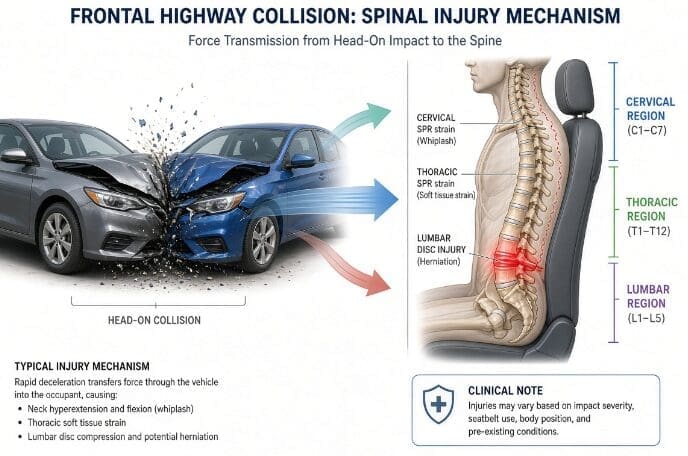
In a frontal collision the vehicle stops abruptly, but the occupant keeps moving forward until something stops her. In a modern car that something is the seatbelt and the airbag. The lap portion of the belt holds the pelvis; the shoulder portion crosses the chest from one shoulder to the opposite hip and catches the upper body. Everything above and between those anchor points keeps moving for a fraction of a second longer. The lower back bends forward over the lap belt. The chest and shoulder are loaded where the belt crosses them. The head and neck, which are not held by anything, whip forward into flexion and then rebound backward into extension. You can read more about this sequence in our article on collision injury dynamics.
Because the impact was at the front left corner rather than straight on, the truck also rotated slightly toward the point of impact. That adds a twisting component to the forces on the spine. Drivers also tend to brace on the steering wheel at the moment of impact, which transmits force up the arms into the wrists, shoulders, and the muscles that connect the shoulder blades to the spine. The airbag, which deploys in milliseconds, can burn or abrade the skin of the forearms and wrists, as happened here.
Head position and the head restraint
She recalled that her head restraint was set low relative to her head. A head restraint works best when its top is level with the top of the head, and it sits close behind it. When it is low, the head can rotate backward over it during the rebound phase, which increases the extension load on the neck. Looking straight ahead, rather than turning, is generally protective because a turned neck concentrates force on one side’s joints. In her case the neck was injured, but less severely than the low back.
Belt loading and the low back
The lowest mobile segments of the lumbar spine sit just above the line of the lap belt. When the upper body jackknifes forward over the belt, those segments are bent forward under compression. That combination of flexion and compression is the classic way a lumbar disc is injured: the front of the disc is squeezed, the soft inner material is pushed backward, and the tough outer ring of the disc can bulge or tear at the back, where the nerve roots run. The disc injury in this case was at the lowest lumbar level, L5-S1, which is where this loading is concentrated.
Vehicle damage and energy transfer
A common question in car crash cases is whether minor vehicle damage rules out injury. The answer is no: modern bumpers are designed to absorb low-speed impacts without visible damage, and people can be hurt in collisions that leave little mark on the car. In this case the question does not arise. A totaled pickup, an airbag deployment and belt and airbag marks on the driver all corroborate a substantial transfer of energy to the vehicle and its occupant. Documenting these details carefully is part of how we show that injuries were caused by a crash.
Why symptoms can build over days
Many people feel shaken but not badly hurt at the scene, and only discover the full extent of their injuries over the next few days. Adrenaline masks pain in the first hours. Injured ligaments and muscles become inflamed over 24 to 72 hours; the muscles around the injured joints tighten protectively; and as the person returns to normal activities (lifting a child, sitting in a car, lying down to sleep) the injured tissues are loaded and begin to hurt. A long car journey soon after a collision, as in this case, often makes things worse. In this patient the back hurt immediately, the neck and shoulder pain followed within a day, and the leg symptoms from the disc appeared weeks later as the disc was loaded by everyday activity.
3. Understanding the injury
Before looking at how she was treated, it helps to understand what was injured. This patient had three main problems: sprains of the ligaments and strains of the muscles along the whole spine and both shoulder girdles; an injury to the lowest lumbar disc with irritation of the nerves nearby; and post-collision headaches with difficulty concentrating.
The anatomy involved
The spine is a column of 24 movable vertebrae (seven in the neck, twelve in the mid back and five in the low back) stacked on the sacrum. Between each pair of vertebrae sits a disc: a tough outer ring of fibers (the annulus) around a softer, gel-like center (the nucleus). At the back of each level, two small facet joints guide motion. Ligaments bind the bones together, and layers of muscle (deep stabilizers close to the spine and larger movers on the surface) control and protect the joints. Nerve roots leave the spinal canal at each level through openings called foramina and travel to the arms and legs. The trapezius, a large flat muscle from the base of the skull across the shoulders and down the mid back, connects the neck and shoulder blades and is commonly strained in collisions.
Sprain, strain and their grades
A sprain is an injury to a ligament; a strain is an injury to a muscle or tendon. Both are graded. A grade 1 injury involves microscopic tearing of fibers with pain and tenderness but no loss of stability. A grade 2 injury involves partial tearing with more pain, swelling, protective spasm and loss of motion. A grade 3 injury is a complete tear. Most collision sprains are grade 1 or 2. They are not trivial: partially torn ligaments heal by forming scar tissue, which is less elastic and less well organized than the original tissue, and the joints they support can remain painful and stiff for months.
What was injured in this patient
- Neck (cervical spine): pain, tenderness and muscle spasm, painful restricted motion in every direction and a headache reproduced by compressing the neck. These findings are typical of a whiplash injury.
- Mid back (thoracic spine) and rib cage: pain below the ribs that made her hold her breath when pressure was applied, and pain spreading outward and down toward the waist. Lying flat on a firm surface made breathing uncomfortable.
- Low back (lumbar spine): the main complaint, rated 6 to 9 out of 10 in the first weeks, worse lying flat and with bending, and later spreading into the buttocks, the backs of the thighs and at times to the feet.
- Both shoulder girdles and trapezius muscles: pain at the front and back of both shoulders and across the top of the shoulders, with the left more tender than the right.
- Chest and wrists: seatbelt bruising and airbag burns at the time of the collision, which settled in the first weeks.
- Head: headaches since the collision, with mild brain fog, later described as trouble concentrating, memory difficulty and occasional trouble finding words.
The disc injury
An MRI of the low back in week 5 of her care showed a small protrusion of the L5-S1 disc toward the center and left, extending less than 3 millimeters, with mild narrowing of the spinal canal at that level. A protrusion is a focal bulge of disc material beyond the normal edge of the disc. Even a small protrusion can irritate the nearby nerve roots, not only by pressing on them but also through the chemical inflammation that disc material provokes. That irritation can cause pain, numbness or tingling in the buttock and leg, which is called radicular pain, or, when nerve function is affected, radiculopathy. Our article on spinal decompression explains how disc injuries are treated conservatively.
The MRI also showed mild drying (desiccation) of the discs and mild wear of the facet joints at several levels, and a common developmental variant: the first segment of the sacrum was not fused to the rest and behaved like an extra lumbar vertebra (lumbarization of S1). These background findings are very common in people with no back pain at all, as we discuss below. A second MRI in week 15, during which she found it difficult to stay still, did not clearly show the protrusion. Small protrusions can become less visible over months as swelling settles, and motion blurs small details, so the two studies are read together and a repeat study was recommended at discharge.
Why the low back took longer than the neck
In this patient the neck settled within the first week of care, while the low back remained the main problem for months. That difference is common after frontal collisions with a seatbelt, and it has a mechanical explanation. The neck was injured by a whip of the head forward and back, a sprain of ligaments and muscles that responds well to restoring motion and calming spasm. The low back was loaded under the lap belt in flexion and compression, the pattern that injures the disc itself. Discs have a poor blood supply, especially in their inner layers, so they heal slowly. They are also loaded by almost everything we do: sitting increases pressure inside the lumbar discs, bending forward increases it further, and lifting while bent and twisted, as when picking up a child from the floor, increases it most of all. An injured disc is therefore aggravated many times a day by ordinary life. Each aggravation can restart inflammation around the nerve roots, which is why her leg symptoms came and went for months. Understanding this helps patients make sense of a recovery in which one region improves quickly and another lags behind.
The shoulder girdle after a belt and steering-wheel injury
Her trapezius and shoulder pain proved to be one of her most persistent problems. Several things load the shoulder girdle in a frontal collision. The shoulder belt crosses one shoulder and the chest and decelerates the upper body abruptly. The hands on the steering wheel transmit force up the arms. And the muscles that hold the shoulder blades against the rib cage, chiefly the trapezius, the rhomboids and the levator scapulae, contract hard to brace the head and arms. Afterward these muscles often stay tight and tender, with trigger points that refer pain into the neck and head. In her case, carrying a young child on the left hip, reaching into a car seat and lifting from the floor kept loading the same muscles, and her shoulders remained more sensitive than her low back by the end of care. Shoulder girdle pain of this kind responds to a combination of manual therapy, strengthening of the muscles that stabilize the shoulder blades, postural work and changes to how daily tasks are done.
Breath-holding and the rib cage
One of her more unusual symptoms was difficulty breathing when lying flat on a firm surface, and a tendency to hold her breath when pressure was applied below her ribs. The ribs attach to the mid back at small joints on either side of each thoracic vertebra, and the muscles between the ribs and along the spine move with every breath. When these joints and muscles are sprained or in spasm, a deep breath stretches injured tissue and hurts, so people instinctively breathe shallowly or hold their breath. Lying flat on a hard surface presses on the same joints. Because the provider is medically trained, other causes of breathing difficulty after a collision, such as rib fracture, lung injury or an asthma flare, were considered as well: she had a known history of asthma, but her breathing symptoms were positional, reproduced by pressure on the mid back and eased by changing position, and her chest X-ray on the day of the collision had shown no fracture. The symptom settled as her mid back improved, and supporting her knees when lying on her back helped in the meantime.
The phases of soft-tissue healing
Injured soft tissue heals in overlapping phases, and good treatment follows them:
- Inflammatory phase (first days to about a week): blood flow increases, swelling develops and pain is highest. Protection, gentle movement and pain control are the priorities.
- Repair or proliferative phase (about 1 to 6 weeks): the body lays down new collagen, initially in a disorganized pattern. Controlled movement helps the new fibers line up along the lines of stress.
- Remodeling phase (about 6 weeks to a year or more): the new tissue is strengthened and reorganized in response to load. This is when progressive exercise matters most.
She arrived at our office five weeks after the collision, at the boundary between repair and remodeling. That is why her care began with pain control and restoring motion but moved, as soon as pain allowed, toward active rehabilitation.
How long recovery usually takes
Most people with a neck or back sprain after a collision improve substantially within the first two to three months, and many recover fully. A sizeable minority, however, still have symptoms six months to a year later. Studies of whiplash-associated disorders show that people with higher pain at the start, more widespread symptoms, headaches and early disability tend to recover more slowly (Carroll 20095). A disc injury with nerve symptoms adds time, because discs heal slowly and are loaded by everyday activity. This patient had several of these features at intake: pain of 8 out of 10 across the whole spine, headaches, difficulty sleeping and limitations in almost every daily activity, followed by leg symptoms. Her seven-month course, with substantial improvement but some residual symptoms, is consistent with that profile. Knowing this from the start helps set realistic expectations and plan care in phases rather than expecting a quick fix.
Sensitization: why pain can outlast the injury
When pain persists for weeks, the nervous system itself can become more sensitive. Nerves in the injured area fire more easily, and the spinal cord and brain begin to amplify signals from that area. This process, called sensitization, explains why light pressure can feel very painful, why pain can spread beyond the original injury and why flare-ups occur with ordinary activities. It does not mean the pain is imaginary; it means the alarm system has been turned up. Movement, graded exercise, good sleep, reassurance and treatments that calm the nervous system, such as acupuncture and massage, help turn it back down.
Headaches and concussion without a head strike
She did not hit her head, yet she had headaches from the day of the collision, together with brain fog, fatigue, trouble concentrating, memory difficulty, irritability and anxiety. Headaches after a collision have two common sources. The first is the neck: injured joints and muscles in the upper neck refer pain into the head (cervicogenic headache), and in her case compressing the neck reproduced her headache. The second is the brain itself. A concussion, or mild traumatic brain injury, does not require a blow to the head; rapid acceleration and deceleration can be enough. Practice guidelines advise clinicians to screen for these symptoms after any significant collision and to refer for specialist evaluation if they persist beyond the usual recovery period (Silverberg and colleagues, 202014).
Factors that made recovery harder
Several features of her life and health did not cause the injury but made it harder to recover from:
- Body composition: a body composition scan placed her body mass index in the obese range, with a high visceral fat level. Obesity is associated with more frequent and more persistent low back pain (Shiri and colleagues, 201013), partly through higher mechanical load on the spine and partly through low-grade inflammation.
- Joint hypermobility: she described herself as unusually flexible. Hypermobile joints rely more on muscles than on ligaments for stability, so when muscles are inhibited by pain, the joints are less well protected.
- Foot mechanics: a foot scan found moderate flattening of the arches under load and an uneven distribution of weight between the left and right feet.
- Daily demands: caring for a young child meant lifting, carrying on one hip and getting down to and up from the floor many times a day, all of which load the low back.
- General health: she had asthma, a history of a pregnancy-related blood sugar problem, and during her care she was being evaluated by her own physician for a possible thyroid problem.
4. Presentation at the first visit
When she arrived at our office, about five weeks after the collision, she completed a detailed online accident questionnaire and a paper pain diagram, and she was examined by the dual-licensed provider. Her story, examination and baseline measurements set the course for everything that followed.
History
She described pain in the neck, the upper, mid and low back and both shoulders, rated 8 out of 10 on an average day, 7 out of 10 at the moment, 9 out of 10 at its worst and 3 out of 10 at its best. The pain was aching, tight and dull, with sudden sharp, shocking episodes. Lying flat on a firm surface made her low back and mid back hurt so much that breathing became difficult; bending her knees helped. Bending forward and carrying her child made it worse. Over-the-counter ibuprofen in high doses helped only partly. She had headaches most days since the collision, with a sense of brain fog, and her sleep was disrupted. Getting up from sitting, driving, sitting or standing for long periods, walking, bathing and household work were all painful, although she could still do them.
Asked about her past, she said she had seen a chiropractor occasionally in previous years, mostly after workouts, and on closer questioning described intermittent back discomfort over the years and joint hyperflexibility. She had not needed imaging, prescription medication or regular treatment for her back in the period before the collision, and she had been fully active. She had asthma and seasonal allergies, took a vitamin and magnesium, did not smoke and drank alcohol only occasionally.
What she wanted from care
Asked what she hoped to achieve, her first goal was simple: to sleep without pain and to lie down without her back seizing. Beyond that she wanted to care for her young child without hurting, to sit and drive comfortably, to return to exercise and to understand what was wrong. Writing these goals down at the start matters. They turn a pain score into a life the patient wants to get back to, they let the team choose treatments that serve those activities, and they give a way to measure progress that means something to the patient. At her re-examinations, progress was recorded against these goals: caring for her family, bending over and sleeping.
Examination
- Observation and palpation: tenderness from the upper neck to the base of the spine and at both shoulders, with tight, guarded muscles along the neck, trapezius, shoulder blades and low back, the left shoulder more tender than the right.
- Range of motion: neck, mid back and low back movement severely reduced and painful in every direction.
- Orthopedic tests: compressing the neck reproduced a mild headache; gentle traction of the neck produced pressure in the palms only. Pressure below the ribs reproduced the pain that made her hold her breath.
- Neurological screen: normal strength in the arms; no leg symptoms at that stage.
- Trial of treatment: a gentle flexion-distraction technique for the low back increased her pain to 7 out of 10 and was stopped, an early sign that her low back would need careful, graded treatment.
Baseline measures
We performed two objective baseline tests at the first visit. A body composition analysis measured muscle, fat and body water, including the ratio of extracellular to total body water, a marker associated with swelling and inflammation (read more about body composition testing). It placed her body fat and visceral fat in the high range and her water ratio slightly above the ideal. A laser foot scan measured how her arches behaved under load and how her weight was shared between her feet: it showed moderate flattening of the arches, a difference in arch height between the two feet and about 12 percent more weight on one side, together with mild forward head posture. Custom foot orthotics and a supportive pillow were recommended. These measurements matter because they identify factors, separate from the injury itself, that affect how fast and how completely a patient recovers.
5. Triage and medical work-up
The first question in any injury case is not “how do we treat this?” but “is it safe to treat this conservatively, and what else needs to be checked?” Because the provider in charge was also a licensed nurse practitioner, they answered that question during the same visit.
Red flags
Red flags are findings that suggest a serious problem such as a fracture, infection, tumor, major nerve compression or a vascular injury. The provider asked about loss of consciousness, vomiting, worsening headache, weakness, loss of bladder or bowel control, numbness in the saddle area, fever, unexplained weight loss and night pain, and reviewed what was known of her emergency department X-rays. None of these red flags was present, and the emergency department had found no fracture. That made conservative care safe to begin, with careful monitoring. The team also requested her emergency department records and imaging.
Imaging decisions
Imaging is not needed for most back pain in the first weeks, and guidelines warn against routine early MRI. It becomes appropriate when pain persists despite good conservative care or when nerve symptoms develop (American College of Radiology Appropriateness Criteria9). In this case, during the first week of care she described a sharp pain running from the tailbone down a leg, followed over the next weeks by numbness and tingling in the legs and feet when sitting and pain to both knees. With leg symptoms and pain still at 7 to 8 out of 10 after weeks of treatment, the provider ordered a lumbar MRI in the first week. It was completed in week 5, after a delay for scheduling and authorization, and confirmed the L5-S1 disc protrusion. A second MRI was performed in week 15 as part of reassessment.
Medical evaluation and medication
She had tried high doses of over-the-counter ibuprofen with limited relief. With the disc injury confirmed and pain limiting her ability to exercise, she was referred to a pain management specialist, who began prescription medication in week 7 and later performed injections. The medication brought her low back pain down from 5 or 6 to about 4 out of 10 and made the leg pain less frequent, which allowed active rehabilitation to begin. Later the provider ordered a compounded topical cream combining a local anesthetic, a nerve-calming medicine and an anti-inflammatory, together with a home electrical stimulation unit, a hot and cold pack, a lumbar support and a topical cooling gel. She preferred long-term, non-drug solutions where possible, so magnesium-based topical products and Epsom salt baths were also discussed and used.
Whole-person screening
Integrated care looks beyond the injured joints. Over the course of her care, the team discussed her sleep and mattress, hydration (with targets for daily water intake), her diet and blood sugar, given a history of a pregnancy-related blood sugar problem and a family history of diabetes, and symptoms such as a fast heart rate, heat intolerance and unusual thirst that her own physician was evaluating as a possible thyroid problem. These issues affect energy, inflammation, muscle function and recovery. Addressing them is part of the functional medicine side of our practice.
Screening for concussion symptoms
Because she had headaches and brain fog from the start, a structured concussion symptom questionnaire was completed in week 8. She reported headaches, fatigue, trouble concentrating, irritability, memory difficulty, anxiety, occasional word-finding difficulty and needing the handrail on stairs. She denied dizziness, blurred vision, sensitivity to light or noise, ringing in the ears and balance problems. This pattern (headache, cognitive and mood symptoms without balance or visual problems) is common after a whiplash-type collision. It was documented so that, if the symptoms persisted, she could be referred for a formal brain injury evaluation.
What else was considered
Part of a medical examination is thinking about what else could explain the symptoms, so that nothing important is missed. In this case the team considered several possibilities alongside the obvious diagnosis of spinal sprain:
- Fracture: a high-energy collision can fracture a vertebra or rib. Her emergency department X-rays showed no fracture, and the later MRI showed no fracture, bone bruise or slippage of one vertebra on another.
- Disc injury with nerve irritation: suggested by pain worse with sitting, bending and lying flat, followed by leg symptoms. Confirmed on the first MRI.
- Sacroiliac joint pain: the joints between the sacrum and the pelvis can cause buttock pain. Compression testing of these joints was negative at discharge.
- Hip problems: hip rotation did not reproduce her pain, but extending the hip did, pointing toward the front of the hip and upper lumbar nerves rather than the hip joint itself.
- Cervical nerve root irritation and thoracic outlet compression: considered when her hands became numb with her arms raised and her thumb and index finger showed reduced sensation at discharge, and part of the reason for the neurology referral.
- Concussion: considered because of headaches and cognitive symptoms despite no head strike, and screened with a structured questionnaire.
- Medical causes of fatigue and heart rate changes: her own physician was evaluating a possible thyroid problem, which can affect energy, muscle function and recovery, and the team asked to see the results.
Thinking through these possibilities did not delay her care. It shaped the questions asked at each visit and the tests ordered when the picture changed.
Outcome measures
Progress was tracked with pain ratings for each region at every visit, recorded both by the patient on a daily form and by the clinician in the chart; with range of motion; with functional goals such as caring for her child, bending and sleeping; with repeat body composition scans; and with formal re-examinations in weeks 7 and 14 and a full neurological examination at discharge. Measuring at every visit allowed the team to see flare-ups early and change the plan.
6. The treatment plan
Her care followed a phased, multimodal plan, the approach recommended by current guidelines for whiplash-associated disorders and low back pain (Bussières and colleagues, 20184; Qaseem and colleagues, 201712). Early care controls pain and restores motion; later care builds strength and independence; and medical care, imaging and specialists are brought in when the clinical picture calls for them.
Phase 1 · Weeks 0 to 10
Calm the pain, restore motion, find the cause
Two to three visits a week. Adjustments, soft-tissue therapy, decompression for the disc, cold and electrical stimulation. Lumbar MRI ordered in week 1 and completed in week 5; pain management from week 7.
Phase 2 · Weeks 10 to 19
Medication on board, rehabilitation begins
Exercise sessions twice a week; chiropractic visits reduced to one or two a week. Trunk, hip and hamstring strength, lifting mechanics for childcare, home program.
Phase 3 · Weeks 19 to 31
Consolidate and hand over
Visits tapered to weekly or less. Gentler instrument adjustments and acupuncture when firm work aggravated the low back; orthopedic spine referral; full neurological examination and neurology referral at discharge.
The phases were a framework, not a fixed schedule. Visit frequency was reduced at each re-examination as her function improved, and it was adjusted around her travel, a one-week office closure and two later breaks. Treatments were added when a new problem appeared, such as decompression for the disc, medication when pain blocked progress and acupuncture when firm manual work became counterproductive, and they were dropped when they stopped helping, as cupping and the roller table on her low back were. This is what guidelines mean by multimodal care: not every treatment at every visit, but the right combination for the patient at that stage, reviewed against her progress. Throughout, the aim was to move responsibility for her recovery steadily from the clinic to her own daily routine, so that by discharge she had the knowledge and the habits to manage flares herself and knew exactly when to come back.
Each treatment below is described with what it is, why this patient needed it and what the evidence shows. The evidence is described honestly: some treatments have strong support, and others, especially passive modalities, are best used as adjuncts within a broader plan.
Coordinating care between visits
Much of the work in integrated care happens between visits. In this case staff chased the MRI authorization when it stalled, requested her emergency records, arranged the pain management, imaging, orthopedic and neurology referrals, ordered home equipment such as an electrical stimulation unit and a lumbar support, and rescheduled visits around a one-week office closure and her travel. Changes to the plan were recorded so that every clinician who saw her the next time knew what had changed and why. For a patient juggling pain, a young child and a busy household, having one team keep track of these threads meant fewer missed steps and fewer repeated explanations.
Chiropractic spinal adjustments
What it is. A spinal adjustment (spinal manipulation) is a controlled, quick movement applied by hand to a joint that is not moving normally, to restore motion and reduce pain. Several techniques were used: a full-spine Diversified technique, a Gonstead approach targeting specific segments, gentle traction of the neck and, later, an Activator instrument that delivers a light, fast impulse.
Why she needed it. Examination at every visit found restricted, painful segments from the upper neck to the pelvis. Adjustments restored motion at these segments and reduced her neck and mid back pain; her neck pain fell from 8 to 1 out of 10 within the first week of care. When firm manual adjustments began to aggravate her low back in the third phase, the team switched to the Activator instrument, which she found much more comfortable.
What the evidence shows. Guidelines recommend spinal manipulation as part of multimodal care for neck pain, whiplash-associated disorders and low back pain (Bussières 20163; Bussières 20184). A systematic review found that spinal manipulation produces modest improvements in pain and function in acute low back pain, with minor, short-lived side effects such as soreness (Paige and colleagues, 201711).
Shoulder and extremity adjustments
What it is. Gentle mobilization and adjustment of the joints of the shoulder girdle: the shoulder joint, the joints between the collarbone and shoulder blade, and the movement of the shoulder blade on the rib cage.
Why she needed it. Both shoulders were restricted and painful from the start, especially the left, where the seatbelt crossed and where she carried her child. The shoulder girdle and trapezius pain proved to be one of her most persistent problems.
What the evidence shows. Treating the shoulder girdle and upper back alongside the neck is part of multimodal care for whiplash-associated disorders, because these regions work as a unit (Bussières 20163).
Spinal decompression
What it is. Motorized decompression gently and rhythmically stretches the spine while the patient lies harnessed on a table, aiming to reduce pressure within the disc and around the nerve roots. Read more on our decompression treatments page.
Why she needed it. Her pain was worst lying flat and bending forward, and she had leg symptoms, a pattern that suggested the disc. Decompression at L5-S1 began in week 1 with a light force, increased gradually and was adjusted to her tolerance throughout; the MRI in week 5 confirmed the disc protrusion. She reported in the middle months that decompression helped “a lot”, but at times later in her care it provoked pain into the leg, and the setting was lowered.
What the evidence shows. The evidence for traction and decompression in low back pain is mixed: some patients report clear relief, but controlled trials have not shown consistent benefit over other care (Chou and colleagues, 20176). For that reason it was used as one part of a broader plan, monitored at every visit and adjusted when it aggravated her.
Passive modalities: cold, traction and electrical stimulation
What it is. Cold packs, a roller table that mobilizes the spine segment by segment, and electrical muscle stimulation applied before or after manual treatment. Later she used a home electrical stimulation unit.
Why she needed it. In the first phase her muscles were in protective spasm and her pain was high. These modalities reduced spasm and pain so that adjustments and exercise could be tolerated. When the roller table began to aggravate her low back, it was limited to the upper and mid back.
What the evidence shows. Passive modalities are adjuncts. Guidelines do not recommend them as stand-alone treatment, but they can make active care more tolerable in the early phase (Qaseem 201712; Bussières 20184). In this case they were reduced as active care increased.
Massage and soft-tissue therapy
What it is. Therapeutic massage, instrument-assisted soft-tissue work (“scraping”), percussive massage, trigger point therapy and, later, cupping.
Why she needed it. Tight, tender muscles in the trapezius, the muscles between the shoulder blades, the low back and the buttocks were a consistent finding. Massage gave her short-term relief at many visits. Cupping helped on the day but left her sorer the next day, so it was stopped; deep pressure with elbows also aggravated her and was avoided.
What the evidence shows. A Cochrane review found that massage provides short-term improvement in pain and function in subacute and chronic low back pain (Furlan and colleagues, 20158). Its effects are modest and short lived on their own, which is why it was combined with exercise.
Acupuncture
What it is. Fine needles placed at specific points to reduce pain and muscle tension, delivered by our acupuncture service.
Why she needed it. In week 22 her low back no longer tolerated firm manual work. Acupuncture was added. At that visit her low back pain was 3 out of 10, down from 6 at the preceding visits, and she felt it helped more over the longer term than on the day. During one session a needle caused a brief twitching of the left leg, a normal nerve response that settled immediately.
What the evidence shows. A large individual-patient-data meta-analysis found acupuncture effective for chronic musculoskeletal pain and headache, with effects that largely persist over a year (Vickers and colleagues, 201815), and the American College of Physicians includes it among recommended treatments for low back pain (Qaseem 201712).
Active rehabilitation and exercise
What it is. Supervised rehabilitation sessions twice a week from week 10: a bike and treadmill warm-up; trunk stability work such as bird dogs; upper back strengthening with cable rows, band pull-aparts and lateral raises; and later hip mobility, lunges, step-ups, sit-to-stands, hip bridges and low-back stretches. A written home program for the neck, shoulders and upper back was added in week 12.
Why she needed it. Her hamstrings and hip muscles were weak, hip-hinge movements (bending at the hips to pick something up) provoked her back, and her flexible joints needed muscular support. The first week was hard, with soreness and a new spot of tightness under the right shoulder blade, but within two weeks she reported better stability, better sleep and lower pain. Her low back reached 3 out of 10 in week 12.
What the evidence shows. Exercise is the best-supported treatment for persistent low back pain and a first-line recommendation in every major guideline (Qaseem 201712; Chou 20176). Learn more about our approach to mobility and flexibility.
Education, posture and lifting coaching
What it is. Practical teaching about sleeping positions, mattress support, sitting, lifting and carrying, hydration, stretching and pacing.
Why she needed it. She carried a toddler on her left hip many times a day, played with him on the floor and slept on a soft mattress. She was taught to support her knees when lying on her back, to switch hips and hold the child close, to use a firmer mattress topper, to stretch after workouts, and to take Epsom salt baths after exercise, which she found helpful.
What the evidence shows. Education and self-management are core elements of every guideline for neck and back pain, and they help prevent recurrence (Bussières 20163; Bussières 20184).
Foot biomechanical assessment
What it is. A laser scan of the feet under load, measuring arch collapse, arch height and the balance of weight between the feet.
Why she needed it. Her scan showed moderate pronation and an uneven load between the feet. The feet are the foundation of posture: uneven support changes how the pelvis and spine are loaded with every step, which is a predisposing factor that affects recovery from a back injury. Custom orthotics were recommended.
What the evidence shows. Assessing biomechanical contributors is part of a complete musculoskeletal evaluation. Orthotic management is well established for foot conditions (Koc and colleagues, 202310), and in back pain it is used as a supportive measure where foot mechanics contribute.
Body composition analysis
What it is. A bioelectrical scan that measures muscle, fat, visceral fat and body water, repeated during care.
Why she needed it. Her body fat, visceral fat and water ratio were all above the ideal range. These are recovery factors linked to back pain and inflammation. Repeat scans showed a modest fall in weight and fat and a slight improvement in the water ratio over her first weeks of care, and they guided conversations about nutrition and activity.
What the evidence shows. Obesity increases both the risk of low back pain and the likelihood that it becomes chronic (Shiri 201013). Measuring body composition makes that risk factor visible and trackable.
Pain management, medication and injections
What it is. Prescription medication and injections from a pain management specialist, and topical prescriptions from our provider.
Why she needed it. After the MRI, her pain was too high to begin exercise. Medication from week 7 lowered it enough to start rehabilitation in week 10. She later received injections, but she decided she did not want further injections and preferred non-drug approaches.
What the evidence shows. Guidelines support medication alongside non-drug care when pain prevents participation in rehabilitation (Qaseem 201712). Injections can relieve radicular pain in the short term and are a reasonable option when it persists.
Specialist referrals
What it is. Referrals to an orthopedic spine surgeon in week 22 for the disc injury, and to a neurologist at discharge.
Why she needed it. Her disc-related pain kept recurring despite good care, and at discharge the examination found new nerve findings. She hoped to avoid surgery but was open to specialist advice. Referral does not mean surgery is planned; it means the right expert reviews the problem.
What the evidence shows. Guidelines advise specialist referral when nerve deficits develop or conservative care fails to resolve disc-related symptoms (Bussières 20184; Hutchins 20219).
7. A closer look
What each examination test shows
| Test | How it is done | What it tells us | In this case |
|---|---|---|---|
| Cervical compression | Gentle downward pressure on the top of the head | Pain from irritated neck joints or nerve roots | Reproduced her headache at intake |
| Cervical distraction | Gentle upward lift of the head | Relief suggests joint or nerve compression | Pressure in the palms only |
| Range of motion | Measured movement in each direction | Stiffness, guarding and recovery over time | Severely reduced at intake; mildly reduced or full in most planes by discharge |
| Muscle strength (0 to 5) | Resisted movement of each muscle group | Weakness can point to a specific nerve root | Normal at intake; hamstring, calf, ankle, finger extensor and triceps weakness at discharge |
| Reflexes | Tapping tendons at the knee, ankle and arm | Reduced reflexes can reflect nerve root or peripheral nerve problems | Absent in all four limbs at discharge; needs comparison with baseline |
| Dermatomal sensation | Light touch along the skin areas supplied by each nerve root | Maps which nerve root may be irritated | Reduced at the right thumb and index finger and the right thigh and foot at discharge |
| Femoral nerve stretch | Extending the hip with the knee bent, lying face down | Irritation of the upper lumbar nerve roots | Pain at the front of the left hip |
| Ely’s test | Bending the knee toward the buttock, lying face down | Hip flexor tightness or lumbar irritation | Positive on the right for low back pain |
| Elevated arm stress test | Arms raised, hands opening and closing | Nerve or vessel compression at the neck and shoulder | Numbness on both sides within 10 to 20 seconds |
| Romberg and finger-to-nose | Balance with eyes closed; coordination | Screens the brain’s balance and coordination systems | Normal |
Most of these tests are part of every chiropractic examination in our office. Repeating them at re-examinations is what allowed the new findings at discharge to be recognized.
The medication approach
Medication was used to make rehabilitation possible, not as a substitute for it. She began with over-the-counter anti-inflammatory medicine, which helped little. After the MRI she was referred for prescription medication and later injections, and our provider ordered a compounded topical cream so that medication could be applied directly to the painful area with less effect on the rest of the body. She told the team clearly that she preferred long-term, non-drug solutions, and her care respected that: magnesium, Epsom salt baths, heat, stretching, acupuncture and exercise carried more of the load as her pain fell. A short-acting opioid had helped her briefly right after the collision, but it was not part of her ongoing plan.
Concussion screening in a patient who did not hit her head
It is easy to assume that a person who did not strike her head cannot have a concussion. The forces in a highway collision can move the brain inside the skull even without impact. Her structured questionnaire in week 8 documented headaches, fatigue, trouble concentrating, memory and word-finding difficulty, irritability and anxiety. These symptoms can also come from neck injury, pain, poor sleep and stress, and in most people they improve over weeks to months. If they persist, the next step is a formal brain injury evaluation by a neurologist, with cognitive testing and, where appropriate, mental health support, as concussion guidelines recommend (Silverberg 202014). Blood tests for brain injury markers were designed to help emergency doctors decide whether a head CT is needed in the first hours after injury (Bazarian and colleagues, 20181); their role months later is still being studied, so they are considered only on a specialist’s advice.
Exercise progression stages
- Protect and mobilize (weeks 0 to 10): gentle movement, knee-supported lying, short walks, positions that ease the back, and stretches taught at the clinic.
- Stabilize (weeks 10 to 14): warm-up on the bike and treadmill, bird dogs and other trunk control exercises, upper back strengthening with bands and cables.
- Strengthen (weeks 14 to 22): squats, lunges and step-ups; hip mobility; stability ball work; hip-hinge training with a dowel; hamstring strengthening.
- Return to function (weeks 22 to 31): continued strength work two days a week, a home program, stretching after workouts and gradual return to longer standing, walking and play with her child.
How flare-ups were managed
Every patient recovering from a spinal injury has flare-ups, and how they are handled matters as much as the overall plan. In this case, flares followed travel, a day of spring cleaning, long days on her feet, the first hard week of exercise, and, later, certain treatments. The approach was the same each time. First, find the trigger: the team asked what she had done in the previous days and which treatments she had received. Second, reduce the load: they lowered the decompression force, replaced firm manual adjustments with the instrument technique, and kept the roller table away from her low back. Third, keep moving: she continued walking and gentle stretching rather than resting in bed, because complete rest tends to prolong pain. Fourth, review the plan: if a treatment aggravated her two visits in a row, she stopped it, as she did with cupping. This approach taught her that a flare isn’t a sign the injury is getting worse or that care has failed but a signal to adjust.
Sleep and recovery
Sleep is when much of tissue repair happens, and pain and poor sleep feed each other. Early in her care, she could not lie flat without pain, and she woke from the pain when she tossed and turned. Later, she found her pain was worse on waking, which she linked to a soft, ageing mattress. Practical changes helped: a pillow or bolster under the knees when lying on her back, a firmer mattress topper rather than an immediate new mattress, a supportive pillow for her forward head posture, a warm bath in the evening and stretching after exercise. She noticed that she slept better on days she exercised, a common and encouraging finding, because better sleep in turn lowers pain sensitivity the next day.
Nutrition, hydration and inflammation
The team discussed hydration from the first weeks, with a practical daily target using her own water bottle, and suggested an electrolyte drink once she was exercising and sweating more. She continued magnesium, which she already took, and used a magnesium-based topical product and Epsom salt baths for muscle tension. Because her body composition scan showed high visceral fat, and because she had a history of a pregnancy-related blood sugar problem, the team talked with her about reducing sugar and refined bread and replacing sweet drinks, changes she had already started. These measures are not a substitute for treating the injury, but visceral fat and blood sugar problems are linked to low-grade inflammation, and addressing them supports healing and reduces the chance that back pain becomes chronic. Our nutrition services are part of the same practice for this reason.
Caring for a young child with a back injury
Parents of young children rarely get to rest an injured back. She lifted and carried a toddler many times a day, often on her left hip, put him in and out of a car seat and played with him on the floor. Each of these tasks loads the low back and the shoulder girdle in exactly the ways that aggravate a disc injury and a strained trapezius. Instead of telling her to avoid them, which was impossible, the team taught her to do them differently: bend at the hips and knees with the child held close, switch hips, kneel on one knee to get down to the floor, use a carrier or stroller for longer outings and avoid twisting while lifting into a car seat. Her rehabilitation exercises, such as hip hinges, lunges, step-ups and sit-to-stands, were chosen because they rehearse these movements under control. By the later months she could go up and down stairs repeatedly and spend a long day on her feet, although a full day of carrying her child could still provoke her back.
What an MRI can and cannot show
MRI is the best test for seeing discs, nerve roots, ligaments and the spinal cord, but it has limits that patients should understand. It shows structure, not pain. Many people without pain have bulging discs and mild joint wear on MRI, so a finding must match the patient’s symptoms and examination before it is considered the cause of pain. MRI also captures one moment: a disc protrusion can shrink as swelling settles, and the inflammation around a nerve root that causes much of the pain from a disc injury may not show at all, as the radiologist on her second study noted. Image quality depends on staying completely still, which is hard for someone in pain, and small findings of two or three millimeters can be lost when the patient moves. Finally, how the levels are counted matters when, as in her case, there is a transitional segment at the base of the spine. For all these reasons, imaging in our practice is always read alongside the history and examination, and when studies disagree, as hers did, the next step is a careful repeat study rather than a conclusion from either one alone.
Hypermobility and rehabilitation
She described herself as unusually flexible, a trait she saw as both a gift and a problem. People with hypermobile joints often enjoy stretching and may feel temporary relief from it, but their joints depend more heavily on muscle control for stability, because their ligaments allow more movement. After an injury, pain inhibits the deep stabilizing muscles, and the joints become vulnerable to small strains with ordinary movements. Rehabilitation for a hypermobile patient therefore emphasizes control over range: slow, precise exercises such as bird dogs and bridges, strength through the middle of the range rather than at its extremes, and careful progression. Adjustments are kept gentle and specific, and aggressive stretching of already mobile segments is avoided. This is one reason the instrument technique suited her better than firm manual adjustments later in her care.
Why several adjustment techniques were used
Chiropractic is not one technique. Diversified technique uses quick manual thrusts across many regions; Gonstead focuses on specific segments identified by palpation and analysis; flexion-distraction gently opens the lower back on a moving table; and the Activator instrument delivers a light, fast impulse with little rotation. Matching the technique to the patient and to the stage of healing is part of good care. Early on, she was treated with Diversified and Gonstead adjustments, which restored motion in her neck and mid back quickly. Flexion-distraction was tried at her first visit but increased her pain, an early sign that her low back was sensitive. Later, when firm adjustments began to aggravate her low back, the Activator kept the benefits of restoring motion without provoking flares. Different clinicians in the team also brought different strengths, and she was encouraged early on to see more than one to find the best fit.
Stress, mood and the injured body
A collision is frightening, and recovery adds stress of its own: pain, poor sleep, the demands of a family, appointments, insurance paperwork and the loss of a vehicle. On her symptom questionnaire she reported irritability and anxiety. Stress and anxiety do not mean pain is “in the head”, but they do increase muscle tension, worsen sleep and amplify pain signals through the same sensitization described earlier. Research on back pain shows that psychological distress is one of the stronger predictors of pain that lasts (Chou and Shekelle, 20107). Integrated care makes room for these factors: explaining the injury clearly, showing progress with measurements, giving practical tools for flares and recommending mental health support or a brain injury evaluation when symptoms persist.
Why documentation matters
Every rating, test and change in the plan was recorded at the visit it happened. That record serves the patient medically, because it shows what helped, what aggravated her and what changed over time, and it is what allows us to recognize a new finding, such as the nerve signs at discharge, as new. It also serves the patient if she needs to explain her injuries to an insurer or in a legal claim. In a car crash case the clinical record is often the most important evidence of how the person was hurt and how she recovered. Our personal injury care is built around careful documentation for that reason.
8. Course of recovery
Her recovery was real but not straight. The summary table and graph below use weeks from her first visit in our office; she came to us about five weeks after the collision.
| Weeks | Phase | Visits | Low back pain | What happened |
|---|---|---|---|---|
| 0 to 2 | Acute and subacute | 5 | 8, then 5 to 7 | Neck pain fell from 8 to 1; headaches eased; decompression started; MRI ordered |
| 2 to 6 | Acute and subacute | 8 | 6 to 8, up to 9 or 10 lying on the table | Leg symptoms appeared; flare after travel; MRI confirmed the disc protrusion; pain management referral; flare after household work |
| 6 to 10 | Medical co-management | 6 | 7.5 falling to 4, flare to 7 | Medication helped; concussion symptom screening; brief flare in week 10 |
| 10 to 14 | Rehabilitation begins | 12 | 4 falling to 3, then 5 | Twice-weekly exercise; sleep and stability improved; re-examination showed progress |
| 14 to 19 | Rehabilitation | 13 | 4 to 5.5, then 3 | Second MRI; one-week office closure; decompression and exercise helping |
| 19 to 23 | Consolidation | 10 | 6 falling to 3 | Flare, then acupuncture and gentle instrument adjustments; orthopedic spine referral |
| 23 to 31 | Taper and discharge | 9 | 3 to 4.5 | Two breaks in care; pain into the leg with decompression; headache flare; final neurological examination and neurology referral |

Weeks 0 to 10: a wide pattern that slowly narrowed
In the first two weeks her neck pain fell quickly, from 8 to 1 out of 10, and her shoulders from 6 to 3. Her low back did not follow. It stayed at 6 to 8 out of 10, and lying flat on the treatment table could push it to 9 or 10. Around week 1 she first noticed sharp pain from the tailbone down a leg; in week 3 numbness and tingling in her feet and legs when sitting; and in week 4 pain to both knees. A long flight in week 5 added to her pain. The MRI that week confirmed the L5-S1 disc protrusion. Medication from the pain management specialist began in week 7, and within two weeks her low back was at 4 out of 10 and the leg pain less frequent. A day of heavy household cleaning in week 6 had already shown how easily ordinary work could provoke her back, and a further flare to 7 in week 10 settled within days.
Weeks 10 to 19: rehabilitation takes hold
Exercise sessions began in week 10. The first week was rough: she was sore, she found “muscles she did not know she had”, and she developed a sharp spot of tightness under the right shoulder blade as the exercises exposed muscle imbalances. Within two weeks the picture had changed. In week 12 she rated her low back and upper back at 3 out of 10, described her stiffness as joint stiffness on waking rather than pain, and said she slept better after workouts. The re-examination in week 14 recorded steady improvement. The office was closed for a week in week 15, and she noticed she did better with two exercise sessions and one adjustment each week.
Weeks 19 to 31: consolidation, flares and a new finding
In week 21 her low back flared to 6. In week 22, after a long day on her feet carrying her child, she still rated it 3, but she was worried that the pain kept returning. Firm adjustments and the roller table were aggravating her low back, so the team switched to the Activator instrument, kept the roller to the upper back and added acupuncture, which she felt helped over the longer term. She was referred to an orthopedic spine surgeon. Two breaks in care followed, of about two and three weeks. When she returned, decompression sometimes provoked pain from the low back to the foot, with tightness in both calves and hamstrings, and the setting was lowered. In week 29 she had a headache rated 7 out of 10 that spread into her neck, shoulders and back. She described a repeating cycle: pain would settle almost to nothing for a day and then return with her daily routine.
What she did between visits
Treatment in the office is only a few hours a week; recovery happens in the other hundred and sixty. Between visits she stretched daily, walked, did her home exercises, took Epsom salt baths after workouts, applied a magnesium-based topical product, used cold packs and a home electrical stimulation unit on bad days, wore a lumbar support for long periods on her feet, supported her knees when lying down and changed how she lifted and carried her child. She also kept the team informed about what helped and what aggravated her, which is how treatments were adjusted. Her comment that two exercise sessions and one adjustment each week worked best for her came from paying attention to her own pattern, and it shaped the schedule for the rest of her care.
Turning points
Looking back over seven months, four moments changed the direction of her care. The first was the appearance of leg symptoms in her first weeks, which led to the MRI and to the diagnosis of a disc injury rather than a simple sprain. The second was the start of prescription medication in week 7, which lowered her pain enough to begin exercise. The third was the start of rehabilitation in week 10: after a hard first week, her stability, sleep and pain improved more in the following month than in the previous two. The fourth was the recognition, in week 22, that firm manual treatment had become counterproductive for her low back, which led to gentler adjustments, acupuncture and an orthopedic referral. Each of these turning points came from listening to her reports and re-examining her, rather than continuing a fixed protocol.
9. Outcome and prognosis
The two body diagrams below summarize her journey. The first shows every area where she reported pain or symptoms during her care. The second shows her presentation at the final examination, as recorded in the provider’s note, with residual areas in red, areas found only on examination hatched, and resolved areas in gray. In both diagrams the front view shows the patient’s left on the reader’s right, the back view shows the patient’s left on the reader’s left, and the region numbers match.


Comparing the two. Of the ten regions in Figure 1, four (the head, the chest, the buttocks and the feet) were no longer reported at the final visit, although headaches had recurred recently. The neck was no longer painful but still restricted in one direction. What remained was the trapezius and shoulder girdle, the mid and low back, and tightness and intermittent pain in the backs of the thighs and calves.
Final findings and what comes next
Her final examination was a full neurological examination by the dual-licensed provider. Strength was normal at the thighs, arms and shoulders, but the hamstrings tested 3 out of 5 on both sides, and the right calf, left ankle, left finger extensors and right triceps tested 4 out of 5. The tendon reflexes at the knees, ankles and elbows were absent on both sides. Light touch was reduced at the right thumb and index finger and along the right thigh and the right L4 and S1 skin areas. Hip extension on the right reproduced her low back pain, stretching the femoral nerve was painful at the front of the left hip, and raising her arms produced numbness in both hands. Balance, coordination and joint position sense were normal.
Some of these findings may reflect irritation of the lumbar and cervical nerve roots. Others, such as symmetrical absent reflexes in all four limbs, can be constitutional in some people and need comparison with a baseline. Either way, they are new compared with her first examination and they need explaining. She was released from active chiropractic care and referred to a neurologist, with nerve conduction and muscle testing, a repeat lumbar MRI, continued physical therapy focused on the hamstrings, calves and core, and a follow-up visit in four to six weeks. If her headaches and concentration problems persist, a formal brain injury evaluation is the next step. If her trapezius and shoulder girdle pain persists despite rehabilitation, regenerative injections such as platelet-rich plasma may be discussed; the evidence for them varies by condition and is strongest for some tendon problems (Ye and colleagues, 202516), so they are an option rather than a routine step.
Why we look for nerve findings at discharge
A discharge visit is easy to treat as a formality: the patient feels better, the visits have tapered and everyone is ready to move on. In our practice the final visit includes a full neurological examination, for three reasons. First, nerve problems can develop slowly during recovery, especially with a disc injury that keeps flaring, and they can be missed if no one tests strength, reflexes and sensation. Second, the findings at discharge are the baseline against which any future change will be judged. Third, a patient should not be told she has recovered when her examination shows something that still needs explaining. In this case the examination found weakness, absent reflexes and reduced sensation that were not present at intake. Some of these findings may prove benign, but they changed the plan from “released” to “released from chiropractic care, with neurology, nerve testing and repeat imaging”.
Living with residual symptoms
Many people who have had a disc injury and a whole-spine sprain live well with some residual symptoms. The goal is not to be free of every twinge but to know what provokes the back, how to settle it and when to seek help. For this patient that means keeping up two strength sessions a week, stretching the hamstrings and hips, using good lifting technique with her child, protecting her sleep, paying attention to body composition and metabolic health, and returning promptly if new leg or arm symptoms, weakness or a change in headaches develop. Supportive visits during flares, tapering over time, help people stay active while their own strength takes over.
Prognosis
Her prognosis for further improvement is fair. In her favor are her age, her engagement with exercise, the improvement in her sleep and stability, and the large reduction in her low back pain from intake. Against her are the duration of her symptoms beyond six months, the nerve component, the wide pain pattern at the start, recurrent flares with ordinary activity, the physical demands of caring for a young child, body composition and hypermobility, and the cognitive and mood symptoms. Research on whiplash shows that higher initial pain and a widespread pattern predict a slower recovery (Carroll and colleagues, 20095), and studies of low back pain show that persistent symptoms, high initial disability and psychological stress predict pain that lasts (Chou and Shekelle, 20107). People with this profile often keep some residual symptoms and benefit from a plan for flares, a maintained exercise habit and prompt reassessment if nerve symptoms change.
10. How we process a case like this
When a patient finishes care after a collision, our work is not done. We compile the case so that the patient, her other doctors and, where there is a claim, her attorney can see exactly what happened and why.
Record compilation and audit
We gather every chart note, intake form, daily pain form, therapy note, imaging report, referral and billing record, and read every page, including handwritten forms. We check the record against itself: dates, names, diagnoses, codes and template entries. Where we find an error, such as a transcription mistake in a dictated note, we correct it by a separate, dated addendum and never by altering the original entry.
Causation and mechanism
We set out the evidence linking the injuries to the collision: the timing of symptoms, the prior history, the objective findings at each level, the imaging and the biomechanics of the crash. We consider other possible causes and explain how pre-existing factors affected recovery, drawing on published research such as the frequency of disc and joint changes on MRI in people without pain (Brinjikji and colleagues, 20152).
A visual course of care
We map the patient’s pain on body diagrams at intake and at discharge and graph her pain ratings visit by visit, so that her course can be understood at a glance, including the flares.
Future care planning
We set out the care the patient is expected to need and the care she may need if certain conditions arise, each tied to her own findings and to published guidelines.
Communicating with the patient’s other providers
A compiled case is also a communication tool. When a patient is referred to a neurologist, a spine surgeon or a pain specialist, that clinician needs a clear summary of the injury, the imaging, the treatments tried, what helped, what aggravated and what the latest examination showed. Our compiled report provides that summary, so the specialist does not have to piece it together from scattered notes and the patient does not have to repeat seven months of history from memory. The same summary helps the patient’s own primary care clinician understand how the injury interacts with her other health issues.
Turning findings into a future care plan
At the end of care we write down what the patient is likely to need next. For this patient the expected care includes a neurology consultation with nerve conduction and muscle testing, a repeat lumbar MRI compared directly with the earlier studies, an orthopedic spine consultation that had been requested during care, a course of physical therapy, a limited number of supportive chiropractic visits tapering toward self-management, medication for flares and a formal re-examination once the specialist results are available. Other care is planned only if certain conditions arise: a neck MRI if the neurologist confirms an arm nerve problem, an epidural injection if leg pain persists and she chooses it, regenerative injections for persistent shoulder girdle pain, a podiatric assessment for orthotics, and a brain injury evaluation and treatment if her headaches and concentration problems persist. Each item is tied to her own findings and to published guidelines, so the plan makes sense to her other doctors and to anyone reviewing her case.
Medical and chiropractic perspectives in one report
Because our provider holds both licenses, the compiled report brings the chiropractic and medical perspectives together: the joint and muscle findings, the neurological examination, the imaging, the medication and the specialist referrals, in one document written by the clinician who directed the care.
11. Questions patients ask
I did not hit my head. Can I still have a concussion?
Yes. Rapid acceleration and deceleration of the head can injure the brain without any impact. Headaches, brain fog, trouble concentrating, memory problems and irritability after a collision should be reported and screened, as they were in this case. Many of these symptoms can also come from neck injury, pain and poor sleep, which is why a careful evaluation matters.
Why did my back pain start but the leg pain come weeks later?
An injured disc can bulge or leak inflammatory material gradually as it is loaded by sitting, bending and lifting. The nerve root next to it may not become irritated until days or weeks later. New leg pain, numbness or tingling is a reason to tell your provider promptly, because it often changes the plan, as it did here when an MRI was ordered.
My MRI shows “degeneration”. Does that mean my pain is from age, not the crash?
Not necessarily. Mild disc drying and joint wear are very common in people in their 20s and 30s who have no pain at all (Brinjikji 20152). What matters is whether you had symptoms and limitations before the collision and how your symptoms and findings changed afterward. A collision can turn silent changes into painful ones.
Why did my second MRI look different from the first?
Small disc protrusions can shrink or become less visible over months as swelling settles, and if you move during the scan the images lose detail. Radiologists also point out that nerve inflammation may not be visible on MRI at all. That is why the two studies are read together with your examination, and why a repeat study may be recommended.
Is spinal decompression right for a disc injury?
It helps some people, especially when pain is worse with sitting and bending, but the research is mixed. It works best as one part of a plan that includes exercise, and it should be adjusted, or stopped, if it aggravates your symptoms.
Why did my provider change from manual adjustments to an instrument?
When firm manual adjustments start to aggravate a sensitive area, a lighter technique such as the Activator instrument can keep the benefits of restoring motion without provoking a flare. Changing technique in response to how you feel is a sign of careful care, not of failure.
Do I really need exercise if adjustments make me feel better?
Yes. Adjustments, massage and other hands-on treatments reduce pain and restore motion, but their effects are temporary. Exercise builds the strength and control that protect your spine in the long run, and it is the best-supported treatment for persistent back pain.
Why did my provider measure my body composition and scan my feet?
Body fat, visceral fat and body water are linked to inflammation and to how long back pain lasts, and the way your feet carry weight affects how your pelvis and spine are loaded. Measuring these factors shows what else needs attention for a full recovery.
What does it mean that I was referred to a neurologist?
It means the examination found changes in your strength, reflexes or sensation that a nerve specialist should evaluate, usually with nerve conduction and muscle tests. It does not mean something terrible is wrong; it means the findings need an expert explanation before your recovery can be called complete.
Will I always have some pain?
Many people recover fully, but people whose symptoms last beyond six months, who have nerve involvement or who had widespread pain at the start are more likely to keep some symptoms. A good exercise habit, a plan for flares and early reassessment if things change give you the best chance of staying well.
12. A practical guide for patients
The first days after a collision
- Get checked the same day, especially if you have chest, neck or back pain, headache, numbness or confusion.
- Keep moving gently; short walks are better than staying in bed.
- Use cold packs for 15 to 20 minutes at a time for new pain and swelling.
- Write down every symptom, including headaches and trouble concentrating, and when each started.
- Avoid long drives in the first days if you can; if you must travel, stop every hour to walk.
The first weeks
- See a provider who can examine your spine and nervous system and order imaging if needed.
- Report new leg or arm pain, numbness, tingling or weakness promptly.
- If lying flat hurts, bend your knees over a pillow or bolster.
- Take medication as directed, and tell your provider if it is not helping.
- Drink enough water, eat regular meals and protect your sleep.
As you improve
- Start the exercise program as soon as your provider advises, and expect some soreness in the first week.
- Stretch after workouts; tight hamstrings and hips load the low back.
- Keep a pain diary so you and your provider can see patterns and triggers.
- Do not stop your home exercises when the pain settles; they are what keeps it away.
Setting up your day
- Change position every 30 to 45 minutes when sitting.
- Use a supportive mattress or a firmer topper, and a pillow that keeps your neck level.
- Consider supportive footwear or orthotics if your feet roll in.
- Warm baths, including Epsom salt baths, can ease muscle tension after activity.
Returning to family, work and recreation
- When lifting a child, bend at the hips and knees, hold the child close and avoid twisting.
- Switch hips when carrying, and use a carrier or stroller for longer outings.
- Get down to and up from the floor using a half-kneel rather than bending at the waist.
- Pace long days on your feet and plan rest breaks.
- Build back up to sports and gym work gradually, with your provider’s guidance.
Driving and travel while you recover
- Adjust the head restraint so its top is level with the top of your head and it sits close behind it.
- Sit with your hips back in the seat and a small support in the curve of your low back.
- On long drives or flights, stand up and walk every hour, and do gentle back and neck movements at stops.
- Expect a flare after a long journey and plan a lighter day afterward.
- If you feel anxious driving after a collision, tell your provider; it is common and help is available.
Questions to ask your provider
- What exactly is injured, and how long does that usually take to heal?
- Do I need imaging now, and what would change your mind?
- Which symptoms should make me call you before my next visit?
- What can I do at home every day to help?
- How will we know if the plan is working, and what happens if it is not?
- Will my strength, reflexes and sensation be checked before I am discharged?
When to seek urgent care
- Loss of bladder or bowel control, or numbness in the groin or inner thighs.
- Rapidly worsening weakness in an arm or leg.
- A severe or sudden headache, repeated vomiting, confusion or drowsiness.
- Chest pain, shortness of breath at rest or fainting.
- Fever with back pain.
13. Ten key takeaways
- A frontal collision loads the whole spine. The belt holds the pelvis and chest while the neck and low back keep moving, which is why this patient was hurt from the neck to the low back and across both shoulders.
- No head strike does not mean no brain symptoms. Her headaches, brain fog and concentration problems were screened with a structured questionnaire and documented for follow-up.
- New leg symptoms change the plan. When pain began running into her legs, an MRI was ordered in her first week of care and confirmed an L5-S1 disc protrusion.
- One provider with two licenses closes gaps. Imaging, medication, specialist referrals and hands-on care were coordinated by the same clinician, without the delays of separate offices.
- Medication can open the door to rehabilitation. Pain management lowered her pain enough to begin exercise, which then did the long-term work.
- Exercise is the backbone of recovery. After a hard first week, rehabilitation improved her stability, sleep and pain, and her low back reached 3 out of 10.
- Good care changes when the patient does. When firm adjustments, the roller table and cupping aggravated her, they were replaced by an instrument technique and acupuncture.
- Whole-person factors shape recovery. Body composition, foot mechanics, hypermobility, childcare lifting and metabolic health were measured and addressed, not ignored.
- Recovery is rarely a straight line. Travel, housework, long days on her feet and treatment changes produced flares that settled, and the pain diary and graph made the pattern visible.
- Discharge is an examination, not a formality. A full neurological examination at her last visit found new weakness, reflex and sensory changes, and she was referred to neurology the same day.
14. Glossary of terms
- Acute, subacute, chronic- Stages of an injury by time: the first weeks, about 6 to 12 weeks, and beyond about 3 months.
- Activator: A handheld instrument that delivers a quick, light adjustment.
- Annulus: The tough outer ring of fibers of a spinal disc.
- Body composition analysis
- A scan that measures muscle, fat, and body water, including the extracellular water ratio.
- Concussion (mild traumatic brain injury)
- A brain injury from a blow or rapid movement of the head, which can cause headaches and cognitive symptoms.
- Decompression—motorized traction that gently stretches the spine to reduce pressure on a disc.
- Dermatome—The skin area supplied by one spinal nerve root.
- Desiccation—Drying of a disc, seen as darkening on MRI.
- Facet joints- The small paired joints at the back of each spinal level.
- Hypermobility—Joints that move more than usual, relying more on muscles for stability.
- L5-S1- The disc between the lowest lumbar vertebra and the sacrum.
- Lumbarization: A common developmental variant in which the first sacral segment behaves like an extra lumbar vertebra.
- Multimodal care
- Care that combines several treatments, such as manual therapy, exercise, education and medication.
- Protrusion A focal bulge of disc material beyond the normal edge of the disc.
- Pronation: Inward rolling and flattening of the foot’s arch under load.
- Radicular pain and radiculopathy
- Pain along the path of an irritated nerve root; radiculopathy adds loss of nerve function such as numbness, weakness or reflex change.
- Red flags- Findings that suggest a serious problem needing urgent investigation.
- Sensitization: Increased sensitivity of the nervous system that amplifies pain signals.
- Sprain and strain Injuries to ligaments (sprain) and to muscles or tendons (strain).
- Trapezius: The large muscle from the base of the skull across the shoulders and down the mid back.
- Whiplash-associated disorder: Neck injury from rapid flexion and extension, as in a collision.
15. Explore more
- Dr. Alex Jimenez: injury and integrated care
- Auto injuries
- Whiplash chiropractic care
- Neck pain
- Spinal decompression
- Rehabilitation and physical therapy
- Board-certified nurse practitioner care
- ChiroMed integrated medicine
- About ChiroMed
- All ChiroMed services
- Sports injuries
- Contact ChiroMed
- Schedule an appointment
16. References
- Bazarian JJ, Biberthaler P, Welch RD, et al. Serum GFAP and UCH-L1 for prediction of absence of intracranial injuries on head CT (ALERT-TBI): a multicentre observational study. Lancet Neurol. 2018;17(9):782-789. doi:10.1016/S1474-4422(18)30231-X
- Brinjikji W, Luetmer PH, Comstock B, et al. Systematic literature review of imaging features of spinal degeneration in asymptomatic populations. AJNR Am J Neuroradiol. 2015;36(4):811-816. doi:10.3174/ajnr.A4173
- Bussières AE, Stewart G, Al-Zoubi F, et al. The treatment of neck pain-associated disorders and whiplash-associated disorders: a clinical practice guideline. J Manipulative Physiol Ther. 2016;39(8):523-564. doi:10.1016/j.jmpt.2016.08.007
- Bussières AE, Stewart G, Al-Zoubi F, et al. Spinal manipulative therapy and other conservative treatments for low back pain: a guideline from the Canadian Chiropractic Guideline Initiative. J Manipulative Physiol Ther. 2018;41(4):265-293. doi:10.1016/j.jmpt.2017.12.004
- Carroll LJ, Holm LW, Hogg-Johnson S, et al. Course and prognostic factors for neck pain in whiplash-associated disorders (WAD). J Manipulative Physiol Ther. 2009;32(2 Suppl):S97-S107. doi:10.1016/j.jmpt.2008.11.014
- Chou R, Deyo R, Friedly J, et al. Nonpharmacologic therapies for low back pain: a systematic review for an American College of Physicians clinical practice guideline. Ann Intern Med. 2017;166(7):493-505. doi:10.7326/M16-2459
- Chou R, Shekelle P. Will this patient develop persistent disabling low back pain? JAMA. 2010;303(13):1295-1302. doi:10.1001/jama.2010.344
- Furlan AD, Giraldo M, Baskwill A, Irvin E, Imamura M. Massage for low-back pain. Cochrane Database Syst Rev. 2015;(9):CD001929. doi:10.1002/14651858.CD001929.pub3
- Hutchins TA, Peckham M, Shah LM, et al. ACR Appropriateness Criteria low back pain: 2021 update. J Am Coll Radiol. 2021;18(11S):S361-S379. doi:10.1016/j.jacr.2021.08.002
- Koc TA Jr, Bise CG, Neville C, et al. Heel pain – plantar fasciitis: revision 2023. J Orthop Sports Phys Ther. 2023;53(12):CPG1-CPG39. doi:10.2519/jospt.2023.0303
- Paige NM, Miake-Lye IM, Booth MS, et al. Association of spinal manipulative therapy with clinical benefit and harm for acute low back pain: systematic review and meta-analysis. JAMA. 2017;317(14):1451-1460. doi:10.1001/jama.2017.3086
- Qaseem A, Wilt TJ, McLean RM, Forciea MA. Noninvasive treatments for acute, subacute, and chronic low back pain: a clinical practice guideline from the American College of Physicians. Ann Intern Med. 2017;166(7):514-530. doi:10.7326/M16-2367
- Shiri R, Karppinen J, Leino-Arjas P, Solovieva S, Viikari-Juntura E. The association between obesity and low back pain: a meta-analysis. Am J Epidemiol. 2010;171(2):135-154. doi:10.1093/aje/kwp356
- Silverberg ND, Iaccarino MA, Panenka WJ, et al. Management of concussion and mild traumatic brain injury: a synthesis of practice guidelines. Arch Phys Med Rehabil. 2020;101(2):382-393. doi:10.1016/j.apmr.2019.10.179
- Vickers AJ, Vertosick EA, Lewith G, et al. Acupuncture for chronic pain: update of an individual patient data meta-analysis. J Pain. 2018;19(5):455-474. doi:10.1016/j.jpain.2017.11.005
- Ye Z, et al. Platelet-rich plasma versus corticosteroid injection for tendinopathy: a meta-analysis. BMC Musculoskelet Disord. 2025;26(1):339. doi:10.1186/s12891-025-08566-3
Post Disclaimer
General Disclaimer, Licenses and Board Certifications *
Professional Scope of Practice *
The information herein on "A frontal highway collision, a low back disc injury and a whole-spine sprain: An integrated chiropractic and medical case" is not intended to replace a one-on-one relationship with a qualified health care professional or licensed physician and is not medical advice. We encourage you to make healthcare decisions based on your research and partnership with a qualified healthcare professional.
Blog Information & Scope Discussions
Welcome to El Paso's Premier Wellness and Injury Care Clinic & Wellness Blog, where Dr. Alex Jimenez, DC, FNP-C, a Multi-State board-certified Family Practice Nurse Practitioner (FNP-BC) and Chiropractor (DC), presents insights on how our multidisciplinary team is dedicated to holistic healing and personalized care. Our practice aligns with evidence-based treatment protocols inspired by integrative medicine principles, similar to those on this site and on our family practice-based chiromed.com site, focusing on naturally restoring health for patients of all ages.
Our areas of multidisciplinary practice include Wellness & Nutrition, Chronic Pain, Personal Injury, Auto Accident Care, Work Injuries, Back Injury, Low Back Pain, Neck Pain, Migraine Headaches, Sports Injuries, Severe Sciatica, Scoliosis, Complex Herniated Discs, Fibromyalgia, Chronic Pain, Complex Injuries, Stress Management, Functional Medicine Treatments, and in-scope care protocols.
Our information scope is multidisciplinary, focusing on musculoskeletal and physical medicine; wellness; contributing etiological viscerosomatic disturbances within clinical presentations; associated somato-visceral reflex clinical dynamics; subluxation complexes; sensitive health issues; and functional medicine articles, topics, and discussions.
We provide and present clinical collaboration with specialists from various disciplines. Each specialist follows their professional scope of practice and licensure jurisdiction. We use functional health & wellness protocols to treat and support care for musculoskeletal injuries or disorders.
Our videos, posts, topics, and insights address clinical matters and issues that directly or indirectly relate to our clinical scope of practice.
Our office has made a reasonable effort to provide supportive citations and has identified relevant research studies that support our posts. We provide copies of supporting research studies upon request to regulatory boards and the public.
For further discussion on how this information relates to specific care plans or treatment protocols, please ask Dr. Alex Jimenez, DC, APRN, FNP-BC, or contact us at 915-850-0900.
We are here to help you and your family.
Blessings
Dr. Alex Jimenez DC, MSACP, APRN, FNP-BC*, CCST, IFMCP, CFMP, ATN
Email: [email protected]
Multidisciplinary Licensing & Board Certifications:
Licensed as a Doctor of Chiropractic (DC) in Texas & New Mexico*
Chiropractic Licenses:
Texas DC License #: TX5807, Verified: TX5807
New Mexico DC License #: NM-DC2182, Verified: NM-DC2182
Nurse Practitioner Licenses:
Texas APRN License #: 1191402, Verified: 1191402 *
New Mexico CNP License #: 90560, Verified 90560
Florida APRN License #: 11043890, Verified: APRN11043890 *
Colorado License #: C-APN.0105610-C-NP, Verified: C-APN.0105610-C-NP
New York License #: N25929, Verified N25929
Georgia APRN License #: GAA-NP005701
Multi-State Advanced Practice Registered Nurse (APRN*) Texas & Multi-States
Multi-state Compact APRN License by Endorsement (43 States)
Compact Status: Multi-State License: Authorized to Practice in 43 States*
Nursing Licensure Compact: Updated Here
DEA Registration: (Drug Enforcement Agency Registered)
All medical (MDs) and family practice providers (FNP-APRN) are registered and licensed to offer various levels of medication.
Verify Providers' DEA Registration Here
License Verification Link: Nursys License Verifier
* Prescriptive Authority Authorized (DEA Registered Providers). Call if Required
Board Certification:
ANCC FNP-BC: Board Certified Nurse Practitioner*
Education:
Graduate with Honors: ICHS: MSN-FNP (Family Nurse Practitioner Program)
Degree Granted. Master's in Family Practice, MSN Diploma (Cum Laude)
Dr. Alex Jimenez, DC, APRN, FNP-BC*, CFMP, IFMCP, ATN, CCST
(Board Certified: Family Practice Nurse Practitioner—Multistate)*
(Licensed Nurse Practitioner & Chiropractor - Multistate)*
Clinical Director
DC & FNP License (Review Above)
Digital Business Card
NPI: 1205907805
Dr. Maria Cardenas, MD
(Board Certified: Internal Medicine)
(Licensed Medical Doctor)
Medical Director, Clinical Director & Collaborative Physician
NPI # 1164426748
MD License #: J2933
Licenses and Board Certifications:
MD: Medical Doctor
DC: Doctor of Chiropractic
APRNP: Advanced Practice Registered Nurse
FNP-BC: Family Practice Specialization (Multi-State Board Certified)
FNP-BC: Family Practice Across Life Span (Neonatal to Geriatrics)
RN: Registered Nurse (Multi-State Compact License)
CFMP: Certified Functional Medicine Provider
MSN-FNP: Master of Science in Family Practice Medicine
MSACP: Master of Science in Advanced Clinical Practice
IFMCP: Institute of Functional Medicine
CCST: Certified Chiropractic Spinal Trauma
ATN: Advanced Translational Neutrogenomics
Family with Primary Care Focus (Family Nurse Practitioner or FNP)
- The Family Nurse Practitioner (FNP) promotes, maintains, and restores health for individuals and families across the lifespan. FNPs also identify health risks, promote wellness, and diagnose and manage acute and chronic illness.
- The FNP focuses on comprehensive primary care, promoting healthy lifestyles for patients across the lifespan in settings such as private practice, physician offices, and community health centers.
Memberships & Associations:
TCA: Texas Chiropractic Association: Member ID: 104311
TNA: Texas Nurse Association: Member ID: 06458222
TNP: Texas Nurse Practitioner Association ID: 2025091511
AANP: American Association of Nurse Practitioners: Member ID: 2198960
ANA: American Nurses Association: Member ID: 06458222 (District TX01)
| Primary Taxonomy | Selected Taxonomy | State | License Number |
|---|---|---|---|
| No | 111N00000X - Chiropractor | NM | DC2182 |
| Yes | 111N00000X - Chiropractor | TX | DC5807 |
| Yes | 363LF0000X - Nurse Practitioner - Family | TX | 1191402 |
| Yes | 363LF0000X - Nurse Practitioner - Family | FL | 11043890 |
| Yes | 363LF0000X - Nurse Practitioner - Family | CO | C-APN.0105610-C-NP |
| Yes | 363LF0000X - Nurse Practitioner - Family | NY | N25929 |
| Yes | 363LF0000X - Nurse Practitioner - Family | NM |
90560 |
| Yes | 363LF0000X - Nurse Practitioner - Family | GA | GAA-NP005701 |
Dr. Alex Jimenez, DC, APRN, FNP-BC*, CFMP, IFMCP, ATN, CCST
(Board Certified: Family Practice Nurse Practitioner—Multistate)*
(Primary Care Across Lifespan—Neonatal / Pediatric / Adult / Geriatrics)
(Licensed Nurse Practitioner & Chiropractor - Multistate)*
Clinical Director
Digital Business Card
NPI: 1205907805
Dr. Maria Cardenas, MD
(Board Certified: Internal Medicine)*
(Licensed Medical Doctor)*
Medical Director, Clinical Director & Collaborative Physician
NPI # 1164426748
MD License #: J2933
📆 Schedule Appointment: Schedule 24/7 (Click Here)