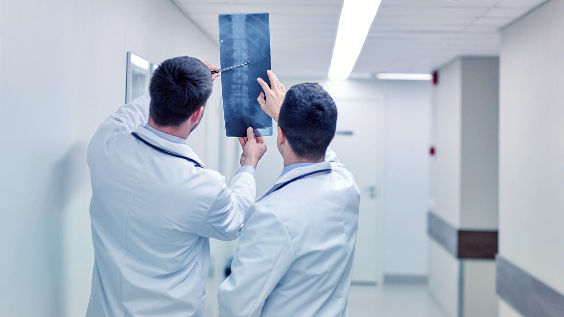
- Conventional Radiography is 2-D imaging modality
- It is required to perform minimum 2-views orthogonal to each other:
- 1 AP (Anterior to Posterior) or PA (Posterior to Anterior)
- 2 Lateral
- Supplemental views: Oblique views etc.
- Skeletal radiographs typically use AP & lateral views
- Chest radiographs and Scoliosis imaging in children will usually use the PA technique
- Exceptions for PA chest views: patients unable to cooperate (severely ill or unconscious patients)
- X-rays are a form of electromagnetic energy (EME) similar to light photons or other sources
- X-rays are a form of man-made radiation
- Ionizing effect of x-rays process of removal of atomic electrons from their orbits
- Two basic types of ionizing radiation:
- Particle (particulate) radiation produced by alpha & beta particles that are the result of radioactive decay of different materials
- Electromagnetic Radiation (EMR) produced by x-rays or gamma rays called photons
- The energy of EMR depends on its wavelength
- Shorter wavelength corresponds to higher energy
- The energy of EME is inversely related to its wavelength
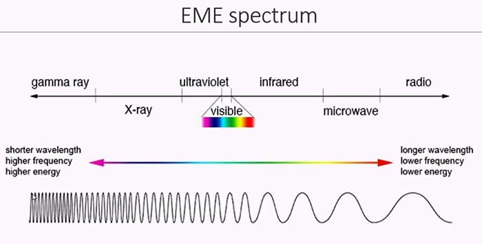

Table of Contents
X-ray Properties
- No charge
- Invisibility
- Penetrability of most matters (esp. human tissues) depends on “Z” (atomic number)
- Making compounds fluoresce and emit light
- Travel at the speed of light
- Ionization and biologic effect on living cells
The Imaging System
- X-rays are produced by an imaging system ( x-ray tube, operator’s console, and high voltage generator)
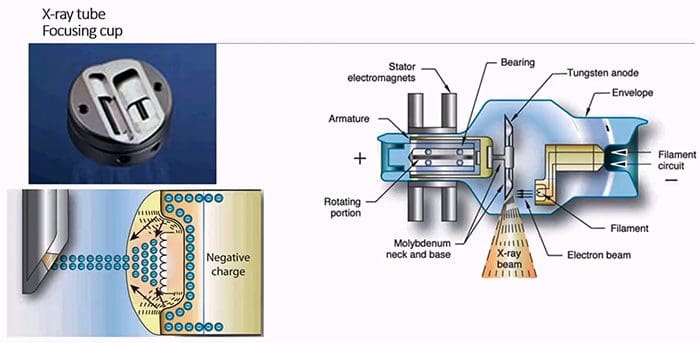
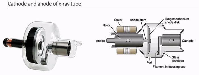
- X-ray tube composed of (-) charged cathode and (+) charged anode enclosed in the evacuated class envelope and housed in the protective coat of metal
- A Cathode made up of filament wire embedded within the focusing cup to give electrostatic focus to electrons’ cloud
- Filament wire of heat resistant thorium tungsten metal of high melting point (3400 C) that “boils off” electrons during thermionic emission
- Focusing cup polished nickel (-) charged that accommodated the filament to electrostatically repulse the electrons and confines them to the focal spot of the anode disc where x-rays are produced
- Anode (+) charged target for electrons to interact at the focal spot
- Conducts electricity
- Rotates to dissipating heat
- Made of tungsten to resist heat
- Anode has a high atomic number to produce x-rays of very high efficiency at the focal spot
- There are 2-focal spots large and small, each corresponding to cathode’s filament size (small vs. large) that depends on the magnitude of current in the cathode dictated by a radiographic study of larger or smaller body parts
- It is known as the dual focus principle
When Electrons are emitted from the cathode as the cloud, they slam into the Anode’s focal spot resulting in 3 man events
- Production of heat (99% outcome)
- Production of Bremsstrahlung (i.e., breaking radiation) x-rays that represent the majority of x-rays within the x-ray emission spectrum
- Production of Characteristic x-rays very few in the emission spectrum
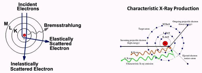
- Newly formed x-rays at the anode are of different energies
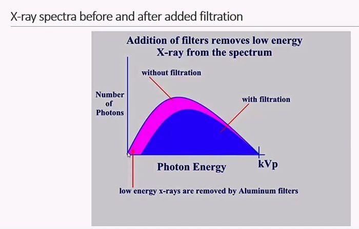
- Only need high energy or “hard” x-rays to perform the radiographic study
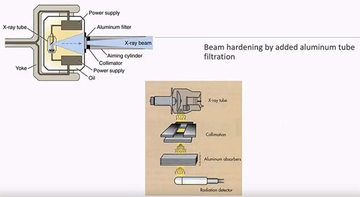
- Before x-rays exiting the tube we need to remove weak or low energy photons, i.e., “harden the beam.”
- Added tube filtration in the form of aluminum filters is used that removes at least 50% of the “unfiltered” beam thus minimizing the patient’s radiation dose and maximizing image quality
High Voltage Generator
- X-ray production requires an uninterrupted flow of electrons to the anode
- Regular electricity supplies AC power with sinusoidal currents of “peaks and drops.”
- In the past, single-phase high voltage generators would convert AC power into a half, or full wave rectified supply with a measure in the thousands of volts delivered with a “voltage ripple” or peaks of high voltage. Therefore, a term kilo voltage peaks (kVp) was used
- Modern generators provide “uninterrupted” flow of electrical potential to the x-ray tube eliminating “voltage ripples” thus referred to as kilovoltage kV without “peaks.”
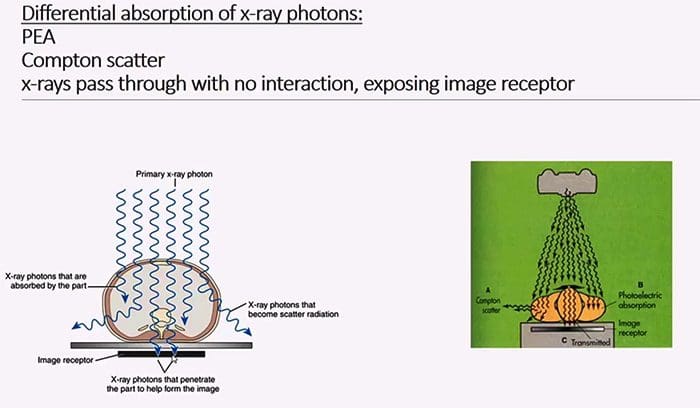
When x-rays interact with the patient’s tissued 3 events will occur
- X-rays will pass through without interaction and “expose” the image receptor
- Photoelectric interaction/effect (PE) comparatively lower energy x-rays will be absorbed/attenuated by the tissues
- Compton scatter x-rays are “bounced off” to form scatter, contributing no useful information to the film and lower image contrast while potentially giving unnecessary radiation dose to staff
- The final image is the product of all three types of interactions known as
- Differential absorption of x-ray photons – the result of photons’ absorption via PE, Compton scatter and x-rays passing through the patient
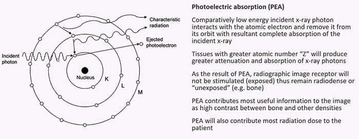
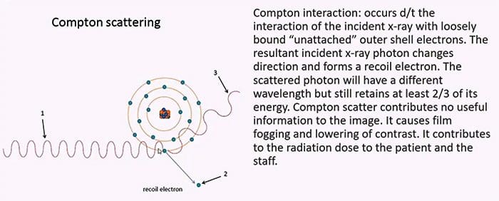
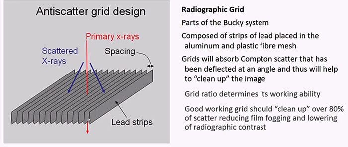
- Compton scatter probability decreases with an increase in x-ray energy compared to PE effect
- Compton effect probability does not depend on the atomic number (Z)
- An increase of total mass density (thick vs. thin) will increase Compton and PE interaction
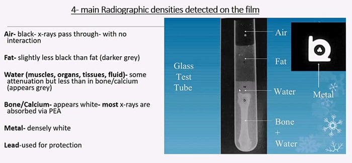
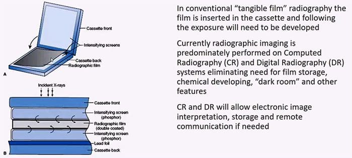
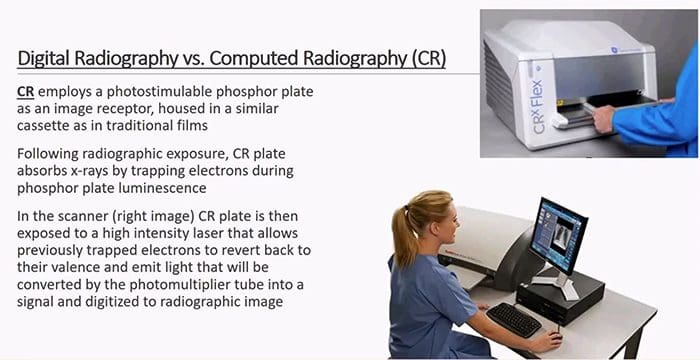
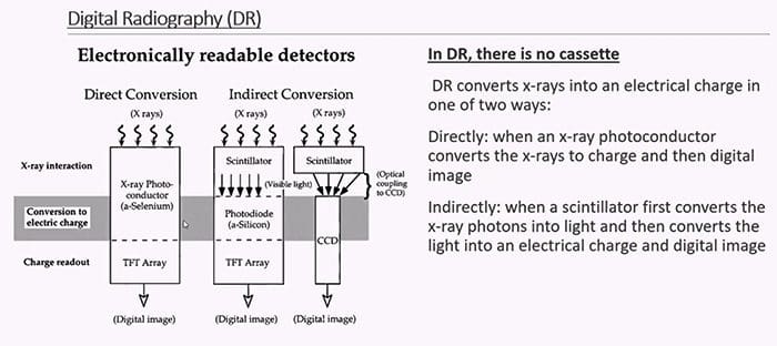
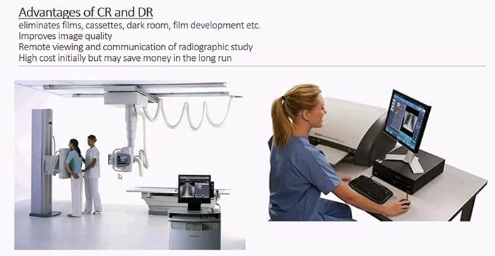
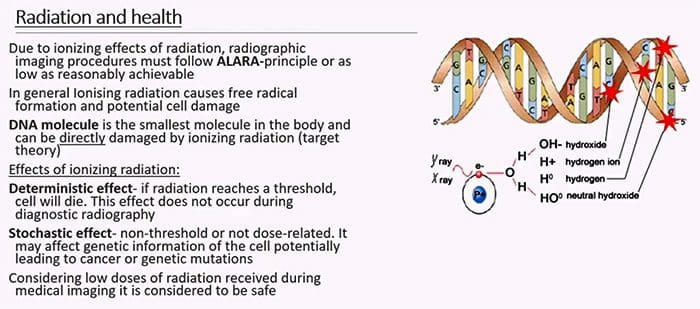
What cells in the body are considered most vulnerable and most resistant to radiation?
- Cells that are rapidly dividing and not terminally differentiated, epithelial cells, etc. are more radiosensitive
- Bone marrow cells (stem cells) & lymphocytes are very radiosensitive
- Muscle & and nerve cells are terminally differentiated and are less sensitive to radiation
- Aged (senescent cells) vs. immature fetal cells are more vulnerable to radiation
- However, following low dose radiation in most healthy individual cells will be able to repair likely without any long-lasting changes

- Pregnancy & radiation initial 6-7 weeks are the most vulnerable
- Do not use routine (non-emergent) radiographic examinations in pregnancy
- Apply 10-day rule establish that radiographs can only be obtained during the initial ten days from the onset of the last menstrual cycle
- Radiographic imaging of children:
- If clinically possible use non-ionizing forms of medical imaging (e.g., ultrasound)

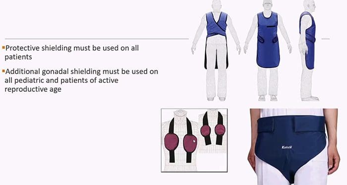
Non-axial imaging studies that use x-ray photons:
- Conventional radiography
- Fluoroscopy
- Mammography
- Radiographic angiography (currently less often used)
- Dental imaging
- Cross-sectional imaging using x-ray photons: Computed Tomography
Indication and Contraindication for conventional radiographic imaging
- Advantages of Radiography: widely available, inexpensive, low radiation burden, the first step in imaging investigation of most MSK complaints
- Disadvantages: 2D imaging, relatively lower diagnostic yield during an examination of soft tissues, numerous artifacts, and dependence on correct radiographic factors selection, etc.
Indications:
- Chest: initial assessment of lung/intrathoracic pathology. Potentially determines or obviates the need for chest CT scanning. Pre-surgical evaluation. Imaging of pediatric patients due to extremely low radiation dose.
- Skeletal: to examine the bone structure and diagnose fractures, dislocation, infection, neoplasms, congenital bone dysplasia, and many forms of arthritis
- Abdomen: can assess acute abdomen, abdominal obstruction, free air or free fluid within the abdominal cavity, nephrolithiasis, evaluate placement of radiopaque tubes/lines, foreign bodies, monitor resolution of postsurgical ileus and others
- Dental: to asses common dental pathologies
Post Disclaimer
Professional Scope of Practice *
The information on this blog site is not intended to replace a one-on-one relationship with a qualified healthcare professional or licensed physician and is not medical advice. We encourage you to make healthcare decisions based on your research and partnership with a qualified healthcare professional.
Blog Information & Scope Discussions
Welcome to El Paso's Premier Wellness and Injury Care Clinic & Wellness Blog, where Dr. Alex Jimenez, DC, FNP-C, a board-certified Family Practice Nurse Practitioner (FNP-BC) and Chiropractor (DC), presents insights on how our team is dedicated to holistic healing and personalized care. Our practice aligns with evidence-based treatment protocols inspired by integrative medicine principles, similar to those found on this site and our family practice-based chiromed.com site, focusing on restoring health naturally for patients of all ages.
Our areas of chiropractic practice include Wellness & Nutrition, Chronic Pain, Personal Injury, Auto Accident Care, Work Injuries, Back Injury, Low Back Pain, Neck Pain, Migraine Headaches, Sports Injuries, Severe Sciatica, Scoliosis, Complex Herniated Discs, Fibromyalgia, Chronic Pain, Complex Injuries, Stress Management, Functional Medicine Treatments, and in-scope care protocols.
Our information scope is limited to chiropractic, musculoskeletal, physical medicine, wellness, contributing etiological viscerosomatic disturbances within clinical presentations, associated somato-visceral reflex clinical dynamics, subluxation complexes, sensitive health issues, and functional medicine articles, topics, and discussions.
We provide and present clinical collaboration with specialists from various disciplines. Each specialist is governed by their professional scope of practice and their jurisdiction of licensure. We use functional health & wellness protocols to treat and support care for the injuries or disorders of the musculoskeletal system.
Our videos, posts, topics, subjects, and insights cover clinical matters and issues that relate to and directly or indirectly support our clinical scope of practice.*
Our office has made a reasonable effort to provide supportive citations and has identified relevant research studies that support our posts. We provide copies of supporting research studies available to regulatory boards and the public upon request.
We understand that we cover matters that require an additional explanation of how they may assist in a particular care plan or treatment protocol; therefore, to discuss the subject matter above further, please feel free to ask Dr. Alex Jimenez, DC, APRN, FNP-BC, or contact us at 915-850-0900.
We are here to help you and your family.
Blessings
Dr. Alex Jimenez DC, MSACP, APRN, FNP-BC*, CCST, IFMCP, CFMP, ATN
email: coach@elpasofunctionalmedicine.com
Licensed as a Doctor of Chiropractic (DC) in Texas & New Mexico*
Texas DC License # TX5807
New Mexico DC License # NM-DC2182
Licensed as a Registered Nurse (RN*) in Texas & Multistate
Texas RN License # 1191402
ANCC FNP-BC: Board Certified Nurse Practitioner*
Compact Status: Multi-State License: Authorized to Practice in 40 States*
Graduate with Honors: ICHS: MSN-FNP (Family Nurse Practitioner Program)
Degree Granted. Master's in Family Practice MSN Diploma (Cum Laude)
Dr. Alex Jimenez, DC, APRN, FNP-BC*, CFMP, IFMCP, ATN, CCST
My Digital Business Card


